


MRI Lumbar Spine Case Study
Our case study of the month is an MRI scan of the lumbar spine. The patient is a male with history of lumbar radiculitis. The MRI Lumbar Spine Case Study procedure included axial and sagittal images of the lumbar spine and they were obtained on our 1.5 Tesla MRI machine and were compared to a previous scan in 2013.
MRI Exam Findings
There is no acute fracture or significant vertebral body lesion. There is a scoliosis, convex to the left in the lumbosacral spine. There is no abnormal signal within the visible spinal cord. There are no intradural abnormalities. There is no paravertebral mass or fluid collection. There are degenerative disc changes at T11-12 and from L1 through S1 in the mild-to-moderate range. At T11-12, there is a small right paracentral protrusion causing minor stenosis. This area was not imaged on the previous examination.
- T12-L1: Normal.There is no disk herniation or evidence of neural compromise.
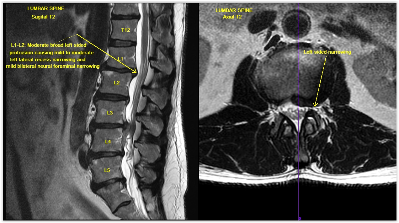
- L1-L2: There is a mild retrolisthesis and moderate broad left-sided protrusion causing mild to moderate left lateral recess narrowing and mild bilateral neural foraminal narrowing, similar to the previous examination.
- L2-L3: There is a slight retrolisthesis, bulge and facet and ligamentum flavum hypertrophy change causing minor stenosis and minor bilateral neural foraminal narrowing without significant change.
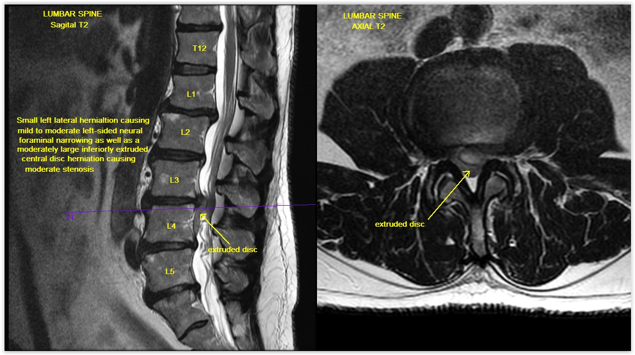
- L3-L4: There is a bulge and facet ligamentum flavum hypertrophy change, similar to the previous examination. However, there is a new small left lateral herniation causing mild to moderate left-sided neural foraminal narrowing as well as a moderately large inferiorly extruded central disc herniation causing moderate stenosis, slightly greater to the left of midline at the L3-4 level with extension inferiorly, behind the superior two thirds of the 4 vertebral body. There is also mild right-sided neural foraminal narrowing at this level.
- L4-L5: There is a slight retrolisthesis, bulge and facet ligamentum flavum hypertrophy change causing minor stenosis and mild bilateral neural foraminal narrowing, similar to the previous examination. L5-S1:There is a bulge and small broad right paracentral protrusion causing minor stenosis and Page: l mild bilateral neural foraminal narrowing without change.
MRI Exam Impression Findings
At L3-4, there is a moderately large inferiorly extruded disc herniation as well as a new small left lateral disc herniation at this same level, as discussed above. There is also multilevel degenerative disease with bulges and protrusions, similar to the previous examination, as discussed above from L1 through S1. A small protrusion at T11-12 is also noted.
For more information on MRI imaging services at Greater Waterbury Imaging Center, visit our clinical section of the website.