

 HISTORY: MULTIPLE BILATERAL RENAL DENSITIES
COMPARISON: None
TECHNIQUE: Multiplanar images of the abdomen were obtained at 1.5 Tesla prior to and following administration of IV contrast.
During this public health emergency, we are using enhanced sterilization processes, social distancing measures and PPE for your protection.
CONTRAST: 18 mL of Dotarem intravenous contrast.
FINDINGS:
Liver: The liver appears normal in size and contour. No mass or intrahepatic biliary dilatation is present. No areas of abnormal enhancement are seen.
Gallbladder: There is metallic artifact in the expected location of the gallbladder neck, consistent with surgical clips. There is a 4.4 x 2.2 x 1.8 cm irregular fluid structure in the gallbladder fossa. There is edema within the adjacent liver parenchyma.
CBD: The common bile duct is dilated measuring up to 10 mm with transition to normal caliber within the pancreatic head near the ampulla.
Pancreas: The pancreas appears normal.
Spleen: The spleen appears normal. A small splenule is present.
Adrenals: The adrenal glands are normal.
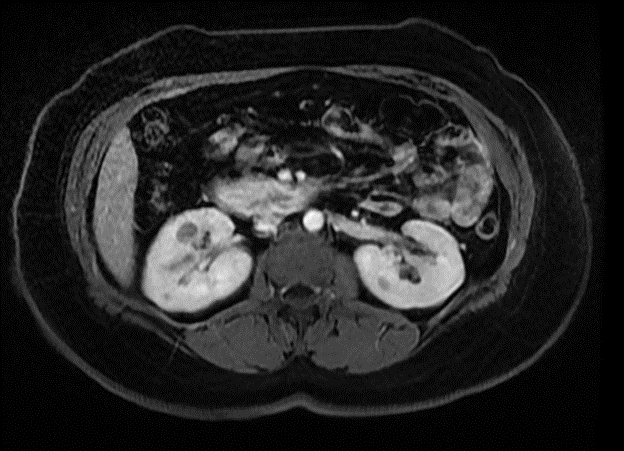
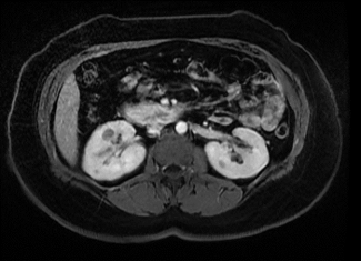
Kidneys: Multiple cystic lesions in both kidneys, the largest is a 1.7 cm lesion in the interpolar region of the right kidney which demonstrates increased intrinsic T1 signal (series 6.2 image 18, series 14 image 37) there is equivocal mild enhancement on the venous and delayed phases, though possibly related to motion artifact. There is an additional 1.1 cm T1 hyperintense lesion in the lower pole of the right kidney (series 14 image 55) also with equivocal appearance on postcontrast venous and delayed phases. The remaining lesions appear predominantly T2 hyperintense without definite enhancement and are most suggestive of simple cysts.
Aorta: No evidence of aortic aneurysm or dissection is seen.
Lymphatics: No abdominal lymphadenopathy is present.
IMPRESSION:
The 1.7 cm cystic lesion in the interpolar right kidney demonstrates increased intrinsic T1 signal and may represent a proteinaceous/hemorrhagic cyst, however, there is equivocal mild enhancement on the venous and delayed phases raising the possibility of hemorrhage associated with an alternate lesion. An additional 1.1 cm lesion in the lower pole of the right kidney has similar characteristics. The presence of multiple additional cysts not demonstrating definite enhancement makes it more likely that the lesions are proteinaceous/hemorrhagic cysts and simple cysts. Follow-up is recommended to ensure benignity, which may consist of renal ultrasound in 3-6 months and/or follow-up MRI abdomen with and without intravenous contrast in 6-12 months.
Irregular fluid structure in the gallbladder fossa measuring 4.4 x 2.2 x 1.8 cm with edema in the adjacent liver is concerning for abscess in the setting of reported prior cholecystectomy, alternatively a postoperative seroma with superimposed inflammation/infection could have this appearance.
Dilated common bile duct, nonspecific though can be seen in the setting of prior cholecystectomy.