
What is Magnetic Resonance Imaging (MRI)?
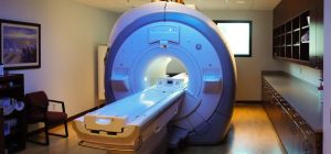
Magnetic Resonance Imaging (MRI) is a non-invasive diagnostic technology that produces detailed images of the internal structures of the body without the use of ionizing radiation. Instead, it utilizes a powerful magnetic field, radio waves, and advanced computer technology to generate clear and precise images of body tissues, including muscles, ligaments, brain tissue, and organs. By aligning the protons in the body to a magnetic field and then using radiofrequency pulses, MRI scans generate cross-sectional images that can be turned into three-dimensional representations. This allows for exceptional visualization of the body’s internal structures and is invaluable in diagnosing a wide range of conditions.
MRI Examinations of the Body: Primarily the Chest, Abdomen, and Pelvis
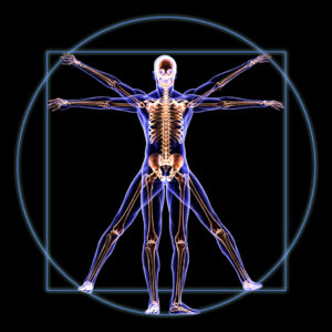
MRI of the body typically encompasses imaging of the chest, abdomen, and pelvis, which are key regions for diagnosing and evaluating numerous diseases and conditions. When performing an MRI of the chest, the focus is often on the heart and lungs, but it can also include the breasts and thoracic spine. An abdominal MRI scans the organs in the abdomen, including the liver, kidneys, pancreas, spleen, and adrenal glands, as well as the gastrointestinal tract.
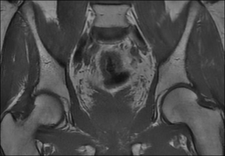
A pelvic MRI provides detailed images of the reproductive organs, bladder, lymph nodes, and bony pelvis. Each of these areas requires specific protocols to optimize the images for the best diagnostic yield.
Common Diagnostic Uses of MR Imaging of the Body
MR imaging of the body serves a multitude of diagnostic purposes. In the chest, it is utilized to investigate abnormalities such as tumors, infections, or diseases of the heart and blood vessels. In the abdomen, it assists in assessing organ function and structure, detecting masses or lesions, and evaluating inflammatory conditions. In the pelvis, MRI is commonly used to examine the reproductive organs for conditions such as fibroids, prostate abnormalities, or pelvic floor disorders. Furthermore, it plays a crucial role in the staging of cancers, guiding biopsies, and planning surgeries.
Specific Types of MR Exams Used for Body Imaging
Magnetic Resonance Imaging (MRI)
This is the standard imaging technique that can be adapted to focus on different body areas for a wide array of clinical indications. It provides a generalized approach that is customizable with various pulse sequences to highlight different tissue characteristics, such as T1-weighted and T2-weighted images for differentiating tissue types and pathologies.
Magnetic Resonance Angiography (MRA)
MRA is a specialized form of MRI that is specifically used to visualize the body’s blood vessels. It provides detailed images of arteries and veins without the need for catheterization or exposure to iodinated contrast materials, which can be advantageous for patients with allergies to these agents. MRA is frequently used to detect aneurysms, blockages, and other vascular abnormalities.
Magnetic Resonance Cholangiopancreatography (MRCP)
MRCP is a specialized type of MRI that targets the biliary and pancreatic ductal systems. It is a non-invasive method to visualize the bile ducts, gallbladder, and pancreatic ducts to identify obstructions, stones, or tumors. This technique is particularly beneficial as it avoids the invasiveness of endoscopic retrograde cholangiopancreatography (ERCP) while still providing high-resolution images.
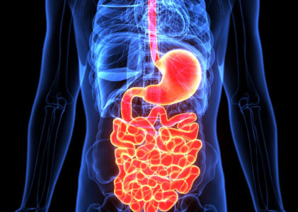
Magnetic Resonance Imaging Enterography (MRE)
MRI Enterography, also known as MRE, is an MRI procedure designed to specifically visualize the small intestine. This is particularly useful in the diagnosis and management of inflammatory bowel diseases, such as Crohn’s disease. It provides detailed images that can reveal inflammation, bleeding, and obstructions within the intestines. Unlike other imaging methods, MRE can provide a comprehensive assessment of the bowel wall and surrounding tissues.
Each of these specific MRI techniques leverages the basic principles of magnetic resonance to visualize structures or functions within the body with exceptional clarity and detail, aiding in the accurate diagnosis and management of a wide range of medical conditions.
MR Examinations for the Diagnosis and Monitoring of Various Conditions
 Tumors of the Chest, Abdomen, or Pelvis
Condition: Tumors within these regions can range from benign growths to malignant cancers. They may originate in the tissues of the chest, such as the lungs or breast, within abdominal organs like the liver or kidneys, or in the reproductive organs in the pelvis.
Use of MRI: MRI is exceptionally useful in imaging tumors due to its high contrast resolution, which differentiates between normal and pathological tissue. For tumors, MRI helps in detection, characterization, and staging. It provides detailed information on the size, extent, and potential spread (metastasis) of tumors, aiding in treatment planning and monitoring response to therapy.
Learn more about MRI for Tumors of the Chest, Abdomen, and Pelvis.
Malformations of the Blood Vessels and Inflammation of the Vessels
Condition: Vascular malformations are congenital abnormalities of the blood vessels that can cause functional issues, while inflammation of the vessels, known as vasculitis, can lead to narrowing or blockage of blood vessels, potentially causing organ damage.
Use of MRA: Magnetic Resonance Angiography (MRA) is a specialized MRI technique that images blood vessels to detect abnormalities such as aneurysms, occlusions, and malformations. MRA is particularly adept at visualizing vessel walls and adjoining soft tissue, making it invaluable for diagnosing vasculitis and for planning surgical or interventional procedures.
Diseases of the Liver
Conditions:
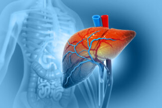
Cirrhosis: A chronic liver disease where normal liver tissue is replaced by scar tissue, affecting liver function.
Abnormalities of the bile ducts and pancreas: These can include blockages, strictures, or congenital variations.
Iron quantifications for genetic hemochromatosis, transfusional hemosiderosis, and chronic hepatopathies: These conditions involve the accumulation of iron in the liver, leading to tissue damage.
Use of MRI: MRI is sensitive to the changes in soft tissue and can detect the early stages of cirrhosis. It also helps in identifying abnormalities within the bile ducts and pancreas. For iron quantification, specialized MRI sequences can quantify liver iron concentration, which is crucial for diagnosing and monitoring conditions such as hemochromatosis and hemosiderosis.
Learn more about MRI for Diseases of the Liver.
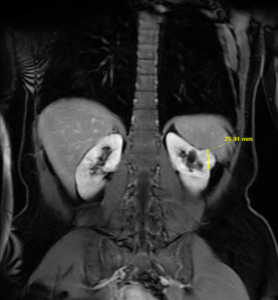 Diseases of the Kidneys
Conditions:
Autosomal dominant polycystic kidney disease: A genetic disorder characterized by the growth of numerous cysts in the kidneys.
Chronic kidney disease: A long-term condition where the kidneys do not function effectively.
Hemorrhagic cyst and cyst infections: Cysts in the kidneys can become infected or bleed.
Tumors: Growth of abnormal tissue in the kidneys.
Use of MRI: MRI provides comprehensive imaging of the kidneys, identifying cysts, tumors, and structural changes associated with chronic kidney disease. It can assess the size and number of cysts in polycystic kidney disease and evaluate complex cysts to determine if they are benign or malignant.
Learn more about MRI for Diseases of the Kidneys.
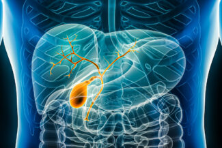
Diseases of the Pancreas and Gallbladder (use MRCP)
Conditions:
Pancreatic cysts: Fluid-filled sacs within the pancreas, which may be benign or precancerous.
Blockages in the ducts: Obstructions in the pancreatic or biliary ducts can cause pain and damage to the liver or pancreas.
Pancreatic cancer staging: Determining the extent and spread of pancreatic cancer.
Gallbladder stones: Hardened deposits that can cause pain and infection.
Use of MRCP: Magnetic Resonance Cholangiopancreatography (MRCP) provides clear images of the pancreatic and biliary ducts, enabling the detection of blockages, stones, and tumors. It is crucial for non-invasively staging pancreatic cancer and guiding the management of pancreatic and biliary diseases.
Learn more about MRI for Diseases of the Pancreas and Gallbladder.
Small Bowel Disease (use MRE)
Conditions:
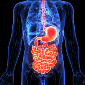
Inflammatory bowel disease (IBD): Crohn’s disease is a type of inflammatory bowel disease (IBD) that can affect any part of the gastrointestinal tract, but most commonly the terminal ileum, which is part of the small bowel.
Use of MRE: Magnetic Resonance Enterography (MRE) is particularly useful for evaluating Crohn’s disease. MRE provides high-resolution images that can show the extent of inflammation, the presence of bowel wall thickening, strictures (narrowed areas), fistulas (abnormal connections to other organs), and abscesses. It is non-invasive and does not involve radiation, making it suitable for repeated use in monitoring the disease progression or response to treatment.
Ulcerative colitis: This is another form of IBD, primarily affecting the colon and rectum. It features continuous inflammation and ulcerations of the colonic mucosa.
Use of MRE: While MRE is not as commonly used for ulcerative colitis as it is for Crohn’s disease, it can still provide valuable information. It can help assess the degree and extent of colonic inflammation, differentiate between ulcerative colitis and Crohn’s disease, and identify complications such as megacolon or colorectal cancer. MRI can also be used to assess the small intestine if involvement beyond the colon is suspected.
Pregnancy
Appendicitis: During pregnancy, inflammation of the appendix can present diagnostic challenges due to the changes in anatomy and the potential risks associated with imaging during pregnancy.
Use of MRI: MRI is used to diagnose appendicitis in pregnant patients because it avoids the ionizing radiation of CT scans. It can effectively visualize the appendix and surrounding tissues to identify signs of inflammation, such as appendiceal enlargement, wall thickening, fat stranding, and fluid collections, which may indicate appendicitis.
Hydronephrosis: This condition is the swelling of a kidney due to a build-up of
urine. It can occur in pregnancy due to the compression of the ureters by the enlarging uterus or the effects of pregnancy hormones relaxing the ureteral muscles.
Use of MRI: MRI can be used safely in pregnancy to assess the extent of hydronephrosis, distinguish it from other causes of abdominal pain, and monitor its progression. It can provide clear images of the renal pelvis, ureter, and bladder to evaluate for obstruction without the use of contrast agents, which are typically avoided during pregnancy.
Fetus in the Womb: Assessing fetal development within the womb is crucial for prenatal care. It includes monitoring the growth, position, and health of the fetus, as well as detecting any congenital abnormalities.
Use of MRI: While ultrasound is the standard imaging modality for fetal evaluation, MRI is used when more detailed information is required, particularly for assessing complex congenital anomalies or when abnormalities are noted on ultrasound that require further clarification. MRI can provide high-resolution images of the fetal brain, spine, thoracic organs, and complex anatomical structures without exposing the fetus to ionizing radiation.
In each of these cases, MRI offers a detailed and safer imaging option compared to other modalities, especially when repeated examinations are necessary or when exposure to ionizing radiation is a concern, such as during pregnancy. The information gleaned from these MRI exams can provide critical insights into the management of the patient’s condition, whether for diagnostic, monitoring, or treatment planning purposes.