This page explores the use of various types of orthopedic MRI of upper extremity, focusing on six specific types of exams: Shoulder MRI, Elbow MRI, Wrist MRI, Brachial Plexus MRI, Humerus and Forearm MRI, and Hand MRI.
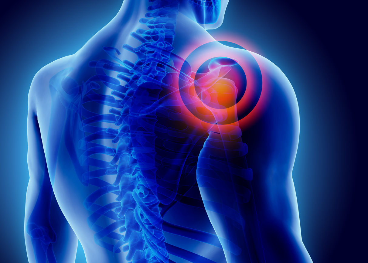
 Shoulder MRI
A Shoulder MRI is instrumental in diagnosing and evaluating a wide range of conditions that affect the shoulder joint and surrounding tissues. Here are the primary indications for a shoulder MRI:
Degenerative Joint Disease
Description: Degenerative joint disease, commonly known as osteoarthritis, involves the wear and tear of the cartilage within the shoulder joint.
MRI Utility: MRI can detect cartilage loss, bone spurs, and changes in the joint’s structure, helping to assess the severity of the disease and guide treatment options.
Arthritis
Description: In addition to osteoarthritis, the shoulder can be affected by rheumatoid arthritis and other inflammatory arthritis.
MRI Utility: MRI helps visualize synovitis, joint effusion, and bone erosions, providing a comprehensive assessment of the inflammatory process and its impact on the joint.
Rotator Cuff Disorders
Description: Rotator cuff disorders, including tears and impingement, are common causes of shoulder pain and dysfunction.
MRI Utility: MRI is highly effective in identifying partial or complete rotator cuff tears, tendinopathy, and impingement syndrome, aiding in precise diagnosis and treatment planning.
Joint Abnormalities
Description: Joint abnormalities such as labral tears, shoulder instability, and dislocations can significantly impact shoulder function.
MRI Utility: MRI provides detailed images of the labrum, joint capsule, and surrounding structures, allowing for accurate diagnosis and management of these conditions.
Infections (e.g., Osteomyelitis)
Description: Infections involving the shoulder, such as osteomyelitis, can cause severe pain and disability if not promptly treated.
MRI Utility: MRI is sensitive in detecting early signs of infection, including bone marrow edema and soft tissue involvement, facilitating early intervention and management.
Tumors Involving Bones and Joints
Description: Tumors, both benign and malignant, can occur in the bones and joints of the shoulder.
MRI Utility: MRI is invaluable in characterizing the extent and nature of bone and joint tumors, guiding biopsy, and planning surgical or other therapeutic interventions.
Pain and Swelling
Description: Persistent pain and swelling in the shoulder can arise from various underlying conditions.
MRI Utility: MRI helps identify the source of pain and swelling, whether it is due to inflammation, injury, or other pathologies, enabling targeted treatment.
Shoulder Pain That Does Not Get Better With Treatment
Description: Chronic shoulder pain unresponsive to conservative treatment requires further investigation.
MRI Utility: MRI provides a thorough evaluation of the shoulder’s internal structures to uncover underlying issues that may have been missed with other imaging modalities.
Decreased Range of Motion
Description: A reduction in the shoulder’s range of motion can result from several conditions, including adhesive capsulitis (frozen shoulder).
MRI Utility: MRI assists in diagnosing conditions that limit shoulder movement by revealing detailed images of the joint and surrounding tissues.
Bicep Tendon Tear
Description: Tears of the bicep tendon can cause significant shoulder pain and weakness.
MRI Utility: MRI effectively identifies the location and extent of bicep tendon tears, crucial for determining the appropriate treatment approach.
Elbow MRI
An Elbow MRI is critical for diagnosing and evaluating a variety of conditions that can affect the elbow joint and surrounding structures. Here are the primary indications for an elbow MRI:
Injuries to the Cartilage and Ligaments
Description: The elbow’s cartilage and ligaments can suffer injuries due to trauma or overuse.
MRI Utility: MRI provides detailed images of the cartilage and ligaments, helping to identify tears, sprains, and other injuries, which is essential for planning appropriate treatment.
Olecranon Bursitis
Description: Olecranon bursitis is inflammation of the bursa located at the tip of the elbow.
MRI Utility: MRI can reveal fluid accumulation and inflammation in the bursa, assisting in the diagnosis and management of bursitis.
Arthritis/Osteoarthritis
Description: Arthritis, including osteoarthritis, can affect the elbow, leading to pain and decreased function.
MRI Utility: MRI is useful for visualizing joint space narrowing, cartilage loss, and other changes associated with arthritis, aiding in diagnosis and treatment planning.
Bicep Tendon Tear
Description: Tears in the bicep tendon at the elbow can cause significant pain and functional impairment.
MRI Utility: MRI effectively identifies the extent and location of bicep tendon tears, which is critical for determining the appropriate treatment strategy.
Tennis/Golf Elbow
Description: Tennis elbow (lateral epicondylitis) and golf elbow (medial epicondylitis) are common overuse injuries affecting the tendons around the elbow.
MRI Utility: MRI can detect tendon degeneration, tears, and inflammation associated with these conditions, helping to guide effective treatment.
Ulnar Nerve Pain
Description: Ulnar nerve entrapment or compression can cause pain, tingling, and weakness in the elbow and forearm.
MRI Utility: MRI can visualize the ulnar nerve and surrounding structures, helping to identify the cause of nerve compression and guiding treatment.
Decreased Range of Motion – Redness, Swelling
Description: Decreased range of motion in the elbow, accompanied by redness and swelling, can result from various underlying conditions.
MRI Utility: MRI assists in diagnosing the causes of decreased range of motion, inflammation, and swelling by providing detailed images of the joint and surrounding tissues.
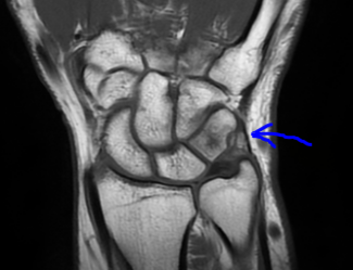 Wrist MRI
A Wrist MRI is essential for diagnosing and evaluating various conditions that affect the complex structure of the wrist. Here are the primary indications for a wrist MRI:
Ligament Tears – Scapholunate Ligament
Description: The scapholunate ligament is a critical stabilizer of the wrist, and tears in this ligament can lead to instability and pain.
MRI Utility: MRI provides high-resolution images that can detect tears and disruptions in the scapholunate ligament, helping to diagnose the extent of injury and plan for appropriate surgical or non-surgical treatments.
Triangular Fibrocartilage Complex (TFCC)
Description: The TFCC is a structure in the wrist that provides stability and support to the distal radioulnar joint and ulnar side of the wrist.
MRI Utility: MRI is highly effective in visualizing injuries to the TFCC, including tears and degenerative changes, which are often the cause of ulnar-sided wrist pain and instability.
Ganglion Cyst
Description: Ganglion cysts are noncancerous lumps that commonly develop along the tendons or joints of the wrist.
MRI Utility: MRI can accurately identify the size, location, and nature of ganglion cysts, aiding in differentiation from other types of masses and guiding treatment options such as aspiration or surgical removal.
Bone Fractures
Description: Bone fractures in the wrist, including scaphoid fractures, can be difficult to detect with standard X-rays.
MRI Utility: MRI is sensitive in detecting occult fractures that are not visible on X-rays, providing detailed images that help in assessing the extent of the fracture and planning appropriate treatment to ensure proper healing.
Inflammation
Description: Inflammation in the wrist can result from various conditions, including tendinitis, tenosynovitis, and inflammatory arthritis.
MRI Utility: MRI can identify and assess the extent of inflammation in the soft tissues, tendons, and joints of the wrist, aiding in the diagnosis of underlying inflammatory conditions and guiding effective treatment strategies.
Carpal Tunnel Syndrome Caused by Median Nerve Enlargement
Description: Carpal tunnel syndrome occurs when the median nerve is compressed as it travels through the carpal tunnel in the wrist.
MRI Utility: MRI can visualize the median nerve and surrounding structures, detecting signs of nerve enlargement, compression, and structural abnormalities that contribute to carpal tunnel syndrome, assisting in both diagnosis and surgical planning if necessary.
Arthritis
Description: Arthritis in the wrist, including rheumatoid arthritis and osteoarthritis, can cause pain, swelling, and decreased function.
MRI Utility: MRI provides detailed images of the joints, cartilage, and soft tissues, allowing for a comprehensive assessment of arthritis-related changes, including joint space narrowing, cartilage loss, and bone erosions.
Kienböck’s Disease
Description: Kienböck’s disease is a condition where the lunate bone in the wrist loses its blood supply, leading to bone death and potential collapse.
MRI Utility: MRI is the gold standard for early detection of Kienböck’s disease, as it can identify changes in the bone marrow indicative of avascular necrosis, helping to diagnose the condition in its early stages and guide appropriate treatment to prevent progression.
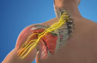
Brachial Plexus MRI
A Brachial Plexus MRI is a specialized MRI of upper extremity imaging technique used to evaluate the complex network of nerves that control the muscles of the shoulder, arm, and hand. Here are the primary indications for a brachial plexus MRI:
Traumatic Compressive or Nontraumatic Plexopathy
Description: Plexopathy refers to damage or disease affecting the brachial plexus. This can result from traumatic injury, compressive forces, or nontraumatic causes such as post-radiation therapy.
MRI Utility: MRI is highly effective in identifying and assessing injuries to the brachial plexus. It can detect nerve damage, inflammation, and structural abnormalities, providing critical information for diagnosing plexopathy and guiding treatment decisions.
Neural Deficit or Muscular Atrophy (Neuritis/Neuropathy)
Description: Conditions such as neuritis (inflammation of the nerves) or neuropathy (nerve damage) can lead to neural deficits and muscular atrophy in the upper extremities.
MRI Utility: MRI provides detailed images of the brachial plexus, helping to identify areas of inflammation, nerve damage, and muscle atrophy. This is essential for diagnosing the underlying causes of neural deficits and planning appropriate therapeutic interventions.
Weakness in the Upper Extremities
Description: Weakness in the upper extremities can result from various conditions affecting the brachial plexus.
MRI Utility: MRI can help determine the cause of weakness by visualizing the brachial plexus and identifying any structural or pathological abnormalities affecting nerve function.
Benign or Malignant Neoplasms
Description: Tumors, whether benign or malignant, can develop in or around the brachial plexus, leading to compression and functional impairment.
MRI Utility: MRI is invaluable in detecting and characterizing neoplasms involving the brachial plexus. It can differentiate between benign and malignant tumors, assess their extent, and guide biopsy and treatment planning.
Compression of the Plexus
Description: Compression of the brachial plexus can occur due to various reasons, including tumors, cysts, or anatomical abnormalities.
MRI Utility: MRI provides detailed images that can identify the source and extent of compression on the brachial plexus, allowing for accurate diagnosis and the development of a targeted treatment plan.
Vascular Abnormalities
Description: Vascular abnormalities, such as aneurysms or vascular malformations, can affect the brachial plexus and surrounding structures.
MRI Utility: MRI, particularly with MR angiography, can visualize vascular structures and identify abnormalities that may impact the brachial plexus. This is crucial for diagnosing vascular conditions and planning appropriate interventions.
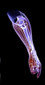
Humerus and Forearm MRI
A Humerus and Forearm MRI is crucial for diagnosing and evaluating various conditions affecting these parts of the upper extremities. Here are the primary indications for a humerus and forearm MRI of upper extremity:
Degenerative Disorders (e.g., Arthritis)
Description: Degenerative disorders such as arthritis involve the deterioration of the joints and surrounding tissues, leading to pain and decreased function.
MRI Utility: MRI provides detailed images of the joints, cartilage, and soft tissues, allowing for a comprehensive assessment of degenerative changes. It helps visualize joint space narrowing, cartilage loss, and bone spurs, aiding in the diagnosis and management of arthritis.
Labral Tears
Description: Labral tears can occur in the shoulder joint where the labrum, a ring of cartilage, helps stabilize the joint.
MRI Utility: MRI is highly effective in detecting labral tears, providing detailed images of the shoulder joint’s soft tissues. This is essential for accurate diagnosis and determining the appropriate treatment, whether surgical or conservative.
Swelling or Bleeding in the Tissues in and Around the Joints
Description: Swelling or bleeding in the tissues around the joints can result from trauma, inflammation, or other underlying conditions.
MRI Utility: MRI can identify and assess the extent of swelling, fluid accumulation, and bleeding within the tissues. This helps in diagnosing the underlying cause and planning effective treatment to reduce inflammation and manage symptoms.
Fractures
Description: Fractures in the humerus or forearm can result from trauma or pathological conditions weakening the bones.
MRI Utility: MRI is sensitive in detecting both acute and occult fractures that might not be visible on X-rays. It provides detailed images of the bone and surrounding soft tissues, aiding in the diagnosis and planning appropriate treatment to ensure proper healing.
Osteomyelitis
Description: Osteomyelitis is an infection of the bone that can cause severe pain, fever, and swelling.
MRI Utility: MRI is the gold standard for early detection of osteomyelitis. It can identify signs of bone infection, including bone marrow edema and abscess formation, facilitating prompt diagnosis and treatment to prevent complications.
Tumors
Description: Tumors, whether benign or malignant, can develop in the bones or soft tissues of the humerus and forearm.
MRI Utility: MRI is invaluable in detecting and characterizing tumors, providing detailed images that help differentiate between benign and malignant growths. It also assesses the extent of the tumor and its impact on surrounding structures, guiding biopsy and treatment planning.
Sprains
Description: Sprains involve the stretching or tearing of ligaments, which can cause pain, swelling, and limited movement.
MRI Utility: MRI provides high-resolution images of ligaments and soft tissues, helping to identify sprains and their severity. This is essential for diagnosing the extent of the injury and planning appropriate treatment to facilitate recovery.
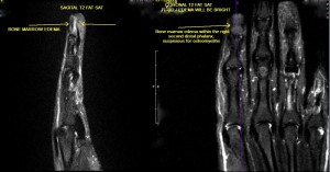 Hand MRI
A Hand MRI is an MRI of upper extremity essential for diagnosing and evaluating various conditions affecting the intricate structures of the hand. Here are the primary indications for a hand MRI:
Fractures
Description: Fractures in the hand bones can result from trauma, falls, or direct impact.
MRI Utility: MRI is sensitive in detecting both acute and occult fractures that might not be visible on X-rays. It provides detailed images of the bones and surrounding soft tissues, helping in the diagnosis and planning appropriate treatment to ensure proper healing and functionality.
Tendon Injuries – Tendonitis, Tendon Tears, and Sheath Infections
Description: Tendon injuries in the hand, such as tendonitis, tendon tears, and infections of the tendon sheath, can cause significant pain and impair movement.
MRI Utility: MRI provides high-resolution images of the tendons and their sheaths, identifying inflammation, tears, and infections. This is essential for accurate diagnosis and determining the appropriate treatment, whether it be conservative management or surgical intervention.
Ligament Tears
Description: Ligament tears in the hand can result from trauma or overuse, leading to pain and instability.
MRI Utility: MRI can detect tears and disruptions in the ligaments of the hand, providing detailed images that help diagnose the extent of injury and plan for appropriate surgical or non-surgical treatments.
Nerve Compression (e.g., Peripheral Neuropathy)
Description: Nerve compression in the hand can cause pain, tingling, numbness, and weakness, often affecting daily activities.
MRI Utility: MRI can visualize the nerves and surrounding structures, identifying areas of compression and abnormalities. This helps in diagnosing conditions like peripheral neuropathy and planning effective treatment strategies to relieve nerve pressure.
Arthritis – Osteoarthritis, Inflammatory, and Rheumatoid
Description: Arthritis in the hand, including osteoarthritis, inflammatory arthritis, and rheumatoid arthritis, can cause joint pain, swelling, and deformity.
MRI Utility: MRI provides detailed images of the joints, cartilage, and soft tissues, allowing for a comprehensive assessment of arthritis-related changes. It helps visualize joint space narrowing, cartilage loss, bone erosions, and inflammation, aiding in the diagnosis and management of various forms of arthritis.
Ganglion Cysts
Description: Ganglion cysts are noncancerous lumps that commonly develop along the tendons or joints of the hand and wrist.
MRI Utility: MRI can accurately identify the size, location, and nature of ganglion cysts, aiding in differentiation from other types of masses and guiding treatment options such as aspiration or surgical removal.
Soft Tissue Infection – Osteomyelitis
Description: Soft tissue infections, including osteomyelitis, can cause severe pain, swelling, and systemic symptoms if not promptly treated.
MRI Utility: MRI is the gold standard for early detection of osteomyelitis and other soft tissue infections. It can identify signs of infection, including bone marrow edema, abscess formation, and soft tissue involvement, facilitating prompt diagnosis and treatment to prevent complications.
Orthopedic MRI of the upper extremities, offers unparalleled detail and accuracy in diagnosing a wide range of conditions. By providing high-resolution images of bones, joints, and soft tissues, MRI helps healthcare professionals develop precise and effective treatment plans, ultimately improving patient outcomes.
This is a detailed overview of the various types of orthopedic MRI exams for the upper extremities. Each section provides critical insights into how MRI technology aids in diagnosing and managing complex conditions, ensuring comprehensive care for patients. Contact GWIC for all your orthopedic imaging needs including an MRI of the upper extremity.