

MRI for Neurological Conditions
What is Magnetic Resonance Imaging (MRI)?
 Magnetic Resonance Imaging (MRI) is a sophisticated imaging technique that uses a powerful magnetic field, radiofrequency pulses, and a computer to produce detailed pictures of organs, soft tissues, bone, and virtually all other internal body structures. MRI does not use ionizing radiation (x-rays). Detailed MR images allow physicians to evaluate various parts of the body and determine the presence of certain diseases that may not be assessed adequately with other imaging methods such as x-rays, ultrasound, or computed tomography (CT).
General Description of Neurological MRI:
Neurological MRI includes comprehensive imaging of the brain and spinal cord—central components of the nervous system. An MRI brain scan focuses on the brain and its surrounding structures to assess for injuries, abnormalities, or diseases. A neuro MRI of the cervical spine examines the neck area for structural abnormalities, tumors, or spinal cord compression. Imaging of the thoracic spine can reveal herniated discs, spinal tumors, or spinal abnormalities. Lastly, an MRI of the lumbar spine assesses the lower back and can detect issues like nerve root compression or lumbar pathology.
Specific Types of MR Exams for Neurological Imaging
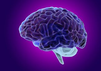
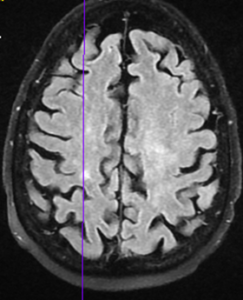
Magnetic Resonance Imaging (MRI) Brain
This exam focuses on the brain to identify conditions such as strokes, tumors, aneurysms, spinal cord injuries, and infections. It can also be used to diagnose causes of headaches and visualize abnormal brain development.
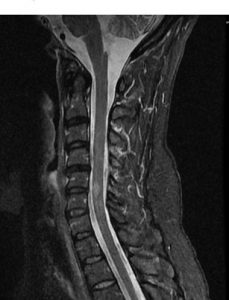
Magnetic Resonance Imaging (MRI) Cervical Spine
An MRI of the cervical spine provides a detailed look at the vertebrae in the neck area, intervertebral discs, spinal cord, and spaces between the vertebrae through which nerves pass. A cervical spine MRI is invaluable in diagnosing herniated discs, spinal stenosis, tumors, and other cervical spine diseases.
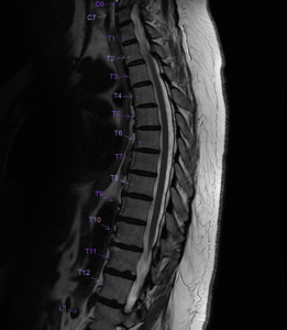
Magnetic Resonance Imaging (MRI) Thoracic Spine
This exam images the mid-back region and is often used to assess abnormalities affecting the thoracic spinal vertebrae and intervertebral discs, such as herniated discs or spinal tumors. It is also used in the evaluation of spinal cord abnormalities.
Magnetic Resonance Imaging (MRI) Lumbar Spine
An MRI of the lumbar spine is used to examine the lower back where many back pain complaints are localized. It can identify disc abnormalities, bone lesions, or tumors in the lumbar spine, and is particularly useful in assessing chronic lower back pain and the causes of sciatica.
Magnetic Resonance Angiography (MRA) Brain
MRA of the brain is a specific type of MRI that focuses on the blood vessels within the brain. It is used to detect, diagnose, and aid the treatment of vascular abnormalities, such as aneurysms, arteriovenous malformations (AVMs), and stenosis of the cerebral arteries.
Magnetic Resonance Angiography (MRA) Neck
MRA of the neck visualizes the cervical blood vessels and can diagnose stenosis, occlusions, dissections, and other abnormalities of the neck arteries and veins. It can be crucial in assessing the vascular supply to the brain and identifying risk factors for stroke.
Magnetic Resonance Imaging (MRI) Pituitary
This exam is specialized for the pituitary gland and surrounding brain structures, often used to investigate disorders related to hormone production. It can detect pituitary tumors, structural abnormalities, and other pathologies affecting the pituitary function.
Magnetic Resonance Imaging (MRI) Internal Auditory Canal

MRI of the internal auditory canal is typically used to assess hearing and balance issues. It can help diagnose acoustic neuromas, multiple sclerosis, and other conditions affecting the cranial nerves associated with hearing and balance.
Neurological MRI of the brain and spine, including specialized exams like MRA, provides critical information for diagnosing and managing neurological conditions. They offer high-resolution images that are vital for accurate diagnosis and treatment planning, aiding healthcare professionals in offering the best patient care.
MRI Help Diagnose and Monitor Many Neurological Conditions
MR examinations are used to help diagnose or monitor treatment for conditions such as:
Blood Clot – MRI Brain
Condition Description: A blood clot in the brain, also known as a cerebral thrombosis or stroke, can block blood flow, leading
to potentially severe neurological deficits. Symptoms may include headache, weakness, confusion, and visual disturbances.
MR Exam Performance: An MRI of the brain is performed to detect the presence and location of a blood clot. It provides detailed images of the brain’s soft tissues and blood vessels, which are critical for identifying the affected areas. MRI with diffusion-weighted imaging (DWI) can detect a clot quickly and with high sensitivity, often within minutes of the event.
Infection – MRI Brain, MRI Cervical/Thoracic/Lumbar Spine
 Condition Description: Infections can affect the brain (encephalitis) and spinal cord (myelitis) or the spaces around them, leading to conditions such as abscesses or meningitis. Symptoms vary but can include fever, pain, neurological deficits, and signs of systemic infection.
MR Exam Performance: MRI exams for infection will target the suspected area—brain or segments of the spine. These scans can detect inflammation, abscess formation, and the spread of infection. MRI with contrast (gadolinium) enhances the visibility of infected areas, as they often light up due to the breakdown of the normal blood-brain barrier.
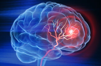
Stroke – MRI Brain, MRA Head, MRA Neck
Condition Description: A stroke occurs when the blood supply to part of the brain is interrupted or reduced, preventing brain tissue from getting oxygen and nutrients. Symptoms include sudden numbness, confusion, trouble speaking, and loss of coordination.
MR Exam Performance: MRI of the brain, along with Magnetic Resonance Angiography (MRA) of the head and neck, is crucial in stroke diagnosis. MRI can show early signs of ischemia, while MRA can evaluate the cerebral arteries for blockages or abnormalities that could lead to a stroke. MRA can also assess the carotid arteries in the neck, which are a common source of emboli that cause strokes.
 Migraines and Headaches – MRI Brain
Condition Description: Migraines are a type of headache characterized by intense, throbbing pain often accompanied by nausea and sensitivity to light and sound. Headaches can have many causes, ranging from tension-type to more serious conditions.
MR Exam Performance: An MRI brain scan for headaches and migraines can rule out structural abnormalities, tumors, stroke, or other brain pathologies that might cause symptoms. While migraines themselves may not be seen on an MRI, the exam is important to exclude other potential causes of the symptoms.
Multiple Sclerosis – MRI Brain, MRI Cervical/Thoracic/Lumbar Spine
Condition Description: Multiple Sclerosis (MS) is an autoimmune disease that affects the central nervous system, leading to demyelination and nerve damage. Symptoms include muscle weakness, balance issues, and fatigue.
MR Exam Performance: MRI is the most sensitive imaging test for MS and is used both for initial diagnosis and monitoring the disease’s progression or response to treatment. It can reveal the presence of lesions or plaques in the white matter of the brain and spinal cord. MRI with contrast can differentiate between active and inactive lesions.
 Dementia and Alzheimer’s – MRI Brain
Condition Description: Dementia is a broad category of brain diseases that cause a long-term decrease in the ability to think and remember. Alzheimer’s disease is the most common cause of dementia and includes symptoms of memory loss, language difficulties, and impulsive or unpredictable behavior.
MR Exam Performance: MRI brain scans in patients with dementia can show loss of brain volume and other structural changes. In Alzheimer’s disease, MRI can detect patterns of brain atrophy that support the diagnosis, particularly in the hippocampal and medial temporal lobe regions.
Hydrocephalus – MRI Brain
Condition Description: Hydrocephalus is characterized by an abnormal accumulation of cerebrospinal fluid (CSF) within the ventricles of the brain, which can lead to increased intracranial pressure and swelling.
MR Exam Performance: MRI is instrumental in diagnosing hydrocephalus. It provides detailed images of the ventricular system and can show enlargement of the ventricles and the flow of CSF, aiding in the determination of the type and severity of hydrocephalus.
Pituitary Gland – MRI Brain Pituitary
Condition Description: The pituitary gland can be affected by various disorders such as tumors, hormonal imbalances, and structural abnormalities.
MR Exam Performance: An MRI focused on the pituitary gland can provide detailed images of the pituitary and the surrounding region of the brain. It is particularly sensitive to detecting small tumors known as adenomas and can assess the effect these tumors may have on adjacent structures.
Traumatic Brain Injury – MRI Brain
Condition Description: Traumatic brain injury (TBI) occurs as a result of a sudden trauma causing damage to the brain. Symptoms can range from mild, such as headaches or temporary confusion, to severe, such as prolonged unconsciousness or amnesia.
MR Exam Performance: MRI is highly sensitive for detecting the subtle changes in brain tissue following a TBI. It can identify microbleeds, diffuse axonal injury, contusions, and brain swelling that may not be visible on a CT scan.
Seizure – MRI Brain
Condition Description: Seizures are sudden, uncontrolled electrical disturbances in the brain that can cause changes in behavior, movements, feelings, and levels of consciousness.
MR Exam Performance: MRI of the brain can identify potential causes of seizures, such as structural brain abnormalities, tumors, stroke, or inflammation. It is especially useful for evaluating the hippocampus for signs of sclerosis, which is common in temporal lobe epilepsy.
Inner Ear Problems – MRI Brain Internal Auditory Canal
Condition Description: Inner ear problems can lead to symptoms like hearing loss, vertigo, and tinnitus. Causes can include benign tumors, such as vestibular schwannomas, or inflammatory conditions.

MR Exam Performance: MRI targeting the internal auditory canal can provide detailed images of the ear’s anatomy and is the best imaging modality for identifying tumors like acoustic neuromas. It can also detect inflammation and other abnormalities affecting the inner ear structures.
Meningitis – MRI Brain
Condition Description: Meningitis is an acute inflammation of the protective membranes covering the brain and spinal cord, known as the meninges. Symptoms include headache, fever, and a stiff neck.
MR Exam Performance: While the initial diagnosis is usually made via lumbar puncture, MRI of the brain can visualize the extent of inflammation and can be particularly helpful in identifying complications such as abscesses, subdural empyemas, and venous sinus thrombosis.
Aneurysm – MRI Brain, MRA Head
Condition Description: An aneurysm i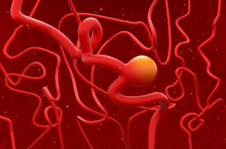 n the brain is a bulge or ballooning in a blood vessel caused by a weakness in the blood vessel wall. If it ruptures, it can lead to a subarachnoid hemorrhage, a stroke, or death.
MR Exam Performance: MRI and MRA of the brain can non-invasively visualize cerebral aneurysms and determine their size, location, and risk of rupture. MRA provides detailed images of the brain’s blood vessels and is particularly adept at identifying aneurysms that might be at risk of bleeding.
In all these conditions, MRI and MRA provide a window into the brain and spinal cord without the risks associated with ionizing radiation, making them indispensable tools in the diagnosis and management of neurological conditions. Contact GWIC for all your MR imaging needs including spine and MRI brain scans.