


NeuroQuant Dementia Case Study
NeuroQuant Dementia Case Study – NeuroQuant is a post-processing software to provide supportive evidence of volume loss in brain disorders through quantitative data.
Modality it uses: Brain MRI on either a 1.5T or 3T MRI scanner
Uses 3D T1, non-contrast, sagittal MRI
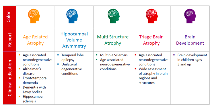
Broad Clinical Assessments: Alzheimer’s disease Epilepsy Multiple Sclerosis (MS) Traumatic Brain Injury (TBI) Brain Development:
Reason for exam: Dementia
Technique: Multiplanar images of the head were obtained at 1.5 Tesla without IV contrast.
Brain: No hemorrhage, mass or infarct. There is a minor pattern of cerebral atrophy diffusely. Minor scattered small vessel ischemic changes are present in the periventricular and subcortical white matter bilaterally. There is a normal pattern of flow within the major arteries at the skull base.
Diffusion images: no evidence of acute ischemic injury.
Sinuses: there is mucosal thickening involving both maxillary sinuses, with a mucus retention cyst filling most of the left maxillary sinus. Mucosal thickening involves the ethmoid air cells bilaterally as well.
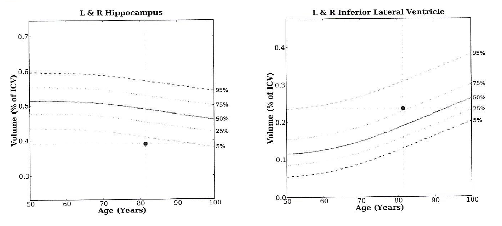
Quantitative volumetric MRI demonstrates relatively low volumes of the left and right hippocampus. The left and right temporal horns of the lateral ventricles are normal in volume, as are the bodies of the lateral ventricles. This pattern of low volume of the hippocami, without ex-vacuo dilation of the temporal horns of the lateral ventricles may be related to early neurodegenerative disease centered on the medial temporal lobes, but follow-up exam would be necessary to determine the trajectory of further volume changes.
Impression: minor atrophy and chronic small vessel ischemic disease. No acute intracranial lesion identified. Low volume hippocampi without ex-vacuo dilatation of the temporal horms of the lateral ventricles, as noted above, may represent early neurodegenerative disease centered on the medial temporal lobes, for which follow-up evaluation would be useful.

Quantitative volumetric MRI demonstrates relatively low volumes of the left and right hippocampus. The left and right temporal horns of the lateral ventricles are normal in volume, as are the bodies of the lateral ventricles. This pattern of low volume of the hippocampi, without ex-vacuo dilatation of the temporal horns of the lateral ventricles may be related to early neurodegenerative disease centered on the medial temporal lobes, but follow-up exam would be necessary to determine the trajectory of further volume changes.
