

Multiple Sclerosis (MS) and MRI: Comprehensive Overview
Table of Contents
Overview of Multiple Sclerosis (MS)

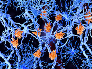
Multiple Sclerosis (MS) is a chronic neurological disorder characterized by inflammation and demyelination of nerve fibers within the central nervous system (CNS). This autoimmune disease leads to the formation of lesions or plaques, disrupting the normal transmission of nerve signals.
MS most commonly affects young adults, particularly those aged 20 to 40, and is more prevalent among women. The disease’s exact cause remains unknown, though genetic predisposition, environmental factors, and immune system anomalies are believed to contribute significantly.
There are several distinct clinical presentations, including:
Relapsing-Remitting MS (RRMS):
- The most common form, affecting approximately 85% of MS patients.
- Characterized by episodes of neurological dysfunction (relapses), such as visual disturbances, numbness, weakness, and balance issues, followed by partial or complete recovery periods (remission).
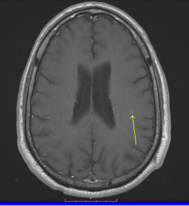
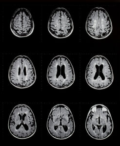
- MRI typically shows multiple lesions that come and go or increase in size and number over time, often involving periventricular, juxtacortical, and infratentorial regions.
- Many RRMS patients eventually transition to SPMS within approximately 10-20 years after onset.
Secondary Progressive MS (SPMS):

- Develops following an initial relapsing-remitting course.
- Marked by a gradual, steady progression of neurological disability, with or without occasional relapses.
- MRI in SPMS typically reveals progressive brain atrophy, fewer new inflammatory lesions, and more stable but extensive chronic lesion burden.
- The transition from RRMS to SPMS signifies a change from inflammatory activity to neurodegenerative processes.
Primary Progressive MS (PPMS):
- Affects about 10-15% of MS patients, with symptoms gradually worsening from the disease’s onset without clear relapses or remissions.
- Common symptoms include mobility issues, steadily increasing weakness, gait impairment, and gradually worsening neurological function.
- MRI typically shows progressive brain and spinal cord atrophy, fewer active inflammatory lesions, and extensive spinal cord involvement, differentiating PPMS from other types.
Clinically Isolated Syndrome (CIS):
- Defined by a single, initial neurological episode lasting at least 24 hours, such as optic neuritis or transverse myelitis, suggestive of MS but not fulfilling diagnostic criteria fully.
- MRI findings can show lesions consistent with demyelination and predict future risk of developing definite MS.
- Approximately 60-80% of patients with CIS and characteristic MRI lesions progress to RRMS within several years, particularly if initial MRI scans reveal numerous lesions.
Symptoms vary significantly across these clinical presentations and may include visual disturbances, motor difficulties, fatigue, cognitive impairment, and sensory abnormalities, substantially impacting patients’ quality of life.
Neurological MRI for Multiple Sclerosis
 Magnetic Resonance Imaging (MRI) is an essential diagnostic tool in neurology, particularly for MS. MRI utilizes powerful magnets, radio waves, and computer technology to generate detailed images of the brain and spinal cord without exposure to radiation.
Common MRI sequences used in MS imaging include:
- T1-weighted imaging: Identifies chronic lesions and brain atrophy.
- T2-weighted imaging: Highlights fluid and inflammation, making lesions clearly visible.
- FLAIR (Fluid-Attenuated Inversion Recovery): Highly sensitive in detecting MS lesions, especially in the brain’s white matter.
- Contrast-enhanced MRI: Uses gadolinium-based contrast agents to detect active inflammation and new lesions.
Typical MS lesions appear primarily in the white matter regions of the brain, spinal cord, and optic nerves. Patients undergoing MRI should understand the procedure’s safety, non-invasive nature, and minimal preparation requirements. GWIC offers advanced imaging with spinal cord MR imaging and MRI brain scans.
How MRI is Used to Diagnose and Monitor Multiple Sclerosis
MRI plays a crucial role in diagnosing MS and differentiating it from other neurological conditions. The McDonald Criteria, regularly updated by international experts, define the diagnostic standards for MS using MRI findings. These criteria require evidence of lesions disseminated in space (multiple CNS locations) and time (new lesions developing over sequential MRIs).
MRI helps track MS progression, document new lesion formation, and gauge treatment efficacy. Patients typically undergo MRI annually, or more frequently during active disease phases or treatment adjustments, aiding clinicians in therapeutic strategies to manage disease progression.
Advancements and Innovations in MRI for MS
Recent advancements in MRI technology significantly enhance MS diagnosis, monitoring, and patient care. These include:
- Diffusion Tensor Imaging (DTI): Visualizes nerve fiber integrity, aiding early detection of subtle neurodegenerative changes.
- Magnetic Resonance Spectroscopy (MRS): Assesses biochemical changes, distinguishing MS from other neurological disorders and evaluating treatment responses.
- Quantitative MRI: Precisely measures lesion volumes, brain atrophy, and spinal cord degeneration, improving disease monitoring accuracy.
- Ultra-high-field MRI (7 Tesla MRI): Offers exceptional resolution to identify smaller lesions and subtle pathologies earlier.
These innovations promise improved clinical management and personalized therapeutic approaches.
Future Outlook for MS
MRI remains essential for MS diagnosis, monitoring, and management. Continued advancements in MRI technology, including AI integration and machine learning, will further enhance diagnostic precision and personalized treatment strategies, significantly benefiting patients and clinicians alike.
Greater Waterbury Imaging Center provides advanced MRI imaging specifically designed for the diagnosis and ongoing monitoring of multiple sclerosis. Our state-of-the-art technology ensures precise detection of lesions and inflammation, helping physicians optimize patient care. Contact us today to schedule your MRI exam and take an active role in managing your MS.