

Table of Contents
Musculoskeletal Magnetic Resonance Imaging for Orthopedic MRI

Welcome to our comprehensive guide on Musculoskeletal Magnetic Resonance Imaging (MRI). Orthopedic MRI advanced imaging techniques play a crucial role in diagnosing and treating orthopedic conditions affecting the musculoskeletal system, including bones, muscles, tendons, ligaments, and soft tissues.
Musculoskeletal MRI is particularly valuable for orthopedic evaluations, providing detailed images that help in the accurate diagnosis of various conditions and guiding effective treatment plans. In this introduction, we will explore the applications and benefits of orthopedic MRI, focusing on the upper extremity, lower extremity, and spine.
What is Orthopedic MRI?
An orthopedic MRI is a non-invasive imaging technique that uses powerful magnets, radio waves, and a computer to create detailed images of the musculoskeletal system. Unlike X-rays and CT scans, MRI does not use ionizing radiation, making it a safer option for patients. The high-resolution images produced by MRI allow for the precise visualization of soft tissues, bone marrow, and other structures, making it an invaluable tool for diagnosing injuries, degenerative diseases, tumors, infections, and congenital anomalies.
Benefits of Musculoskeletal MRI
An orthopedic MRI of the musculoskeletal system provides many benefits including:
- Detailed Imaging: Provides high-resolution images of soft tissues, bones, and joints.
- Non-Invasive: No ionizing radiation, making it safer for repeated use.
- Comprehensive: Can assess multiple structures in a single scan.
- Accurate Diagnosis: Helps in diagnosing a wide range of conditions with high accuracy.
- Guided Treatment: Assists in planning surgeries, guiding biopsies, and monitoring treatment progress.
Orthopedic Upper Extremity MRI
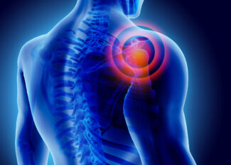
Orthopedic Upper Extremity MRI focuses on the shoulder, elbow, wrist, and hand. This imaging technique is essential for diagnosing conditions such as rotator cuff tears, labral tears, tendonitis, ligament injuries, fractures, and arthritis. It is also useful in detecting soft tissue masses and evaluating post-surgical changes.
Applications:
- Shoulder: Rotator cuff tears, labral tears, bursitis, tendonitis, arthritis, and impingement syndrome.
- Elbow: Tendonitis, ligament injuries, fractures, and joint instability.
- Wrist and Hand: Carpal tunnel syndrome, ligament injuries, fractures, tendonitis, and arthritis.
Common Conditions Diagnosed:
- Rotator Cuff Tears: MRI can accurately visualize tears in the rotator cuff, guiding treatment decisions such as surgery or physical therapy.
- Tendonitis: Inflammation of tendons can be effectively diagnosed, aiding in the implementation of appropriate treatments.
- Arthritis: MRI helps in assessing the extent of arthritis and planning suitable interventions.
Orthopedic Lower Extremity MRI
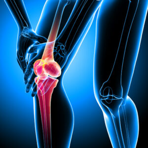
Orthopedic Lower Extremity MRI focuses on the hip, knee, ankle, and foot. This imaging technique is vital for diagnosing injuries and conditions affecting the lower extremities, including ligament tears, meniscal injuries, stress fractures, and degenerative diseases.
Applications:
- Hip: Labral tears, arthritis, fractures, and soft tissue injuries.
- Knee: Meniscal tears, ligament injuries (ACL, PCL, MCL, LCL), cartilage damage, and arthritis.
- Ankle and Foot: Ligament injuries, fractures, tendonitis, and plantar fasciitis.
Common Conditions Diagnosed:
- Meniscal Tears: MRI is highly effective in detecting tears in the meniscus, crucial for deciding between surgical and non-surgical treatments.
- Ligament Injuries: ACL and other ligament injuries are accurately diagnosed, aiding in the appropriate management and rehabilitation strategies.
- Stress Fractures: Early detection of stress fractures helps in preventing further injury and ensuring proper healing.
Orthopedic Spine MRI
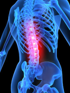
Orthopedic MRI of the spine focuses on the cervical, thoracic, and lumbar spine. This imaging technique is essential for diagnosing spinal conditions such as disc herniation, spinal stenosis, tumors, infections, and degenerative disc disease. It provides detailed images of the vertebrae, intervertebral discs, spinal cord, and surrounding soft tissues.
Applications:
- Cervical Spine: Disc herniation, spinal stenosis, fractures, and degenerative diseases.
- Thoracic Spine: Disc herniation, tumors, infections, and fractures.
- Lumbar Spine: Disc herniation, spinal stenosis, fractures, and degenerative diseases.
Common Conditions Diagnosed:
- Disc Herniation: MRI can pinpoint the location and severity of herniated discs, guiding treatment options such as physical therapy or surgery.
- Spinal Stenosis: Narrowing of the spinal canal can be accurately assessed, helping in the planning of interventions to relieve symptoms.
- Degenerative Disc Disease: MRI helps in evaluating the extent of disc degeneration and formulating appropriate treatment plans.
GWIC Offers Orthopedic MRI Services
Musculoskeletal MRI is an invaluable tool in the field of orthopedics, providing detailed and accurate images that facilitate the diagnosis and treatment of various conditions affecting the musculoskeletal system. Whether it is the upper extremity, lower extremity, or spine, MRI offers critical insights that guide effective patient care.
If you have any questions or need further information about musculoskeletal MRI, please feel free to contact our team of specialists. We are committed to providing the highest quality of care and ensuring the best possible outcomes for our patients with all MR imaging services, including orthopedic MRI.