Table of Contents
Comprehensive Guide to Breast MRI
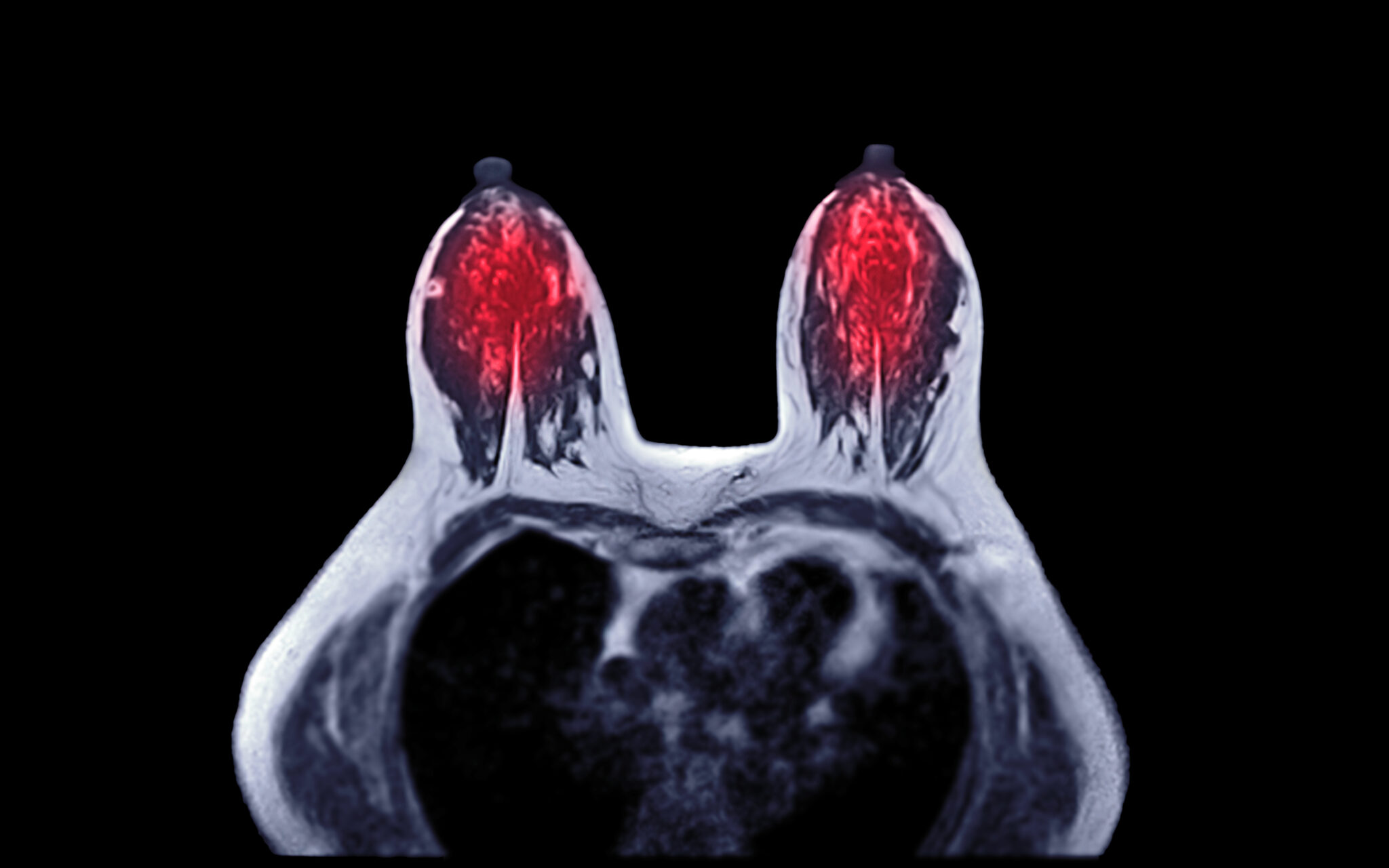
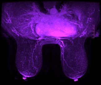
Breast Magnetic Resonance Imaging (MRI) is a non-invasive diagnostic tool that uses powerful magnetic fields and radio waves to create detailed images of the breast tissue. Unlike traditional imaging methods, Breast MRI provides high-contrast, three-dimensional images, allowing for the detection of abnormalities that might be missed by mammography or ultrasound. Since its development in the 1980s, Breast MRI has become an invaluable asset in the early detection and management of breast diseases.
Importance of Breast Imaging in Healthcare
 Breast imaging plays a crucial role in the early detection of breast cancer and other breast-related conditions. Early diagnosis significantly improves treatment outcomes and survival rates. Breast MRI enhances the capabilities of traditional imaging techniques, offering superior sensitivity in detecting invasive cancers, especially in women with dense breast tissue or those at high risk.
This guide is designed for healthcare professionals—including radiologists and women’s healthcare providers—and patients seeking comprehensive information about Breast MRI. It aims to enhance understanding of the technology, its applications, and its benefits, facilitating informed decision-making in clinical practice and personal health management.
Understanding Breast MRI
What is a Breast MRI?
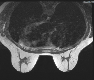
Breast MRI is an advanced imaging technique specifically tailored to evaluate breast tissue. It utilizes magnetic resonance principles to generate detailed images without the use of ionizing radiation. The procedure involves placing the patient inside a large magnet where radiofrequency pulses stimulate hydrogen atoms in the body, producing signals converted into images by a computer.
How Breast MRI Works
The MRI machine generates a strong magnetic field that aligns the protons in hydrogen atoms within the body’s water molecules. Radiofrequency pulses then disrupt this alignment, and as the protons return to their original state, they emit signals captured by the machine. Contrast agents, typically gadolinium-based, are often administered intravenously to enhance image clarity by highlighting abnormal tissue vascularity.
Comparison with Other Imaging Modalities
While mammography and ultrasound are standard breast imaging techniques, they have limitations, especially in dense breast tissue. Mammography uses low-dose X-rays to produce images but may miss small tumors. Ultrasound utilizes sound waves to detect abnormalities but is operator-dependent. Breast MRI surpasses these methods in sensitivity, particularly for detecting invasive cancers and assessing the extent of disease.
Types of Breast MRI Exams
Diagnostic Breast MRI
Diagnostic Breast MRI is performed when there is a need to further investigate abnormalities detected by other imaging methods or physical examination. Indications include evaluating the extent of known cancer, investigating inconclusive findings, and assessing implant integrity.
Screening Breast MRI
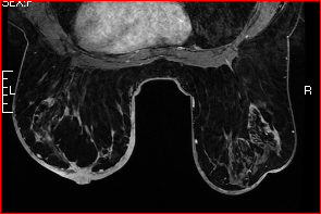
Screening Breast MRI is recommended for individuals at high risk of breast cancer, such as those with a strong family history or genetic predisposition (e.g., BRCA mutation carriers). It is used alongside mammography to increase the likelihood of early detection. Screening frequency is typically annual but may vary based on individual risk factors.
Preoperative Breast MRI
Preoperative Breast MRI assists in surgical planning by accurately delineating tumor size, location, and multifocality. It helps surgeons determine the most appropriate surgical approach, whether breast-conserving surgery or mastectomy, and reduces the likelihood of re-operation.
Advanced and Emerging MRI Techniques
Emerging techniques like Functional MRI and Diffusion-Weighted Imaging (DWI) provide additional information about tumor biology and cellular density. These methods enhance lesion characterization, aiding in distinguishing benign from malignant lesions without additional invasive procedures.
Conditions and Diseases Evaluated with Breast MRI
 Breast Cancer Detection and Staging
Breast MRI is highly sensitive in detecting both invasive and non-invasive cancers, such as ductal carcinoma in situ (DCIS). It plays a pivotal role in staging by assessing tumor size, chest wall involvement, and lymph node status, which are critical for treatment planning.
Evaluation of Breast Implants
For patients with silicone or saline breast implants, MRI is the preferred modality to assess implant integrity. It effectively detects ruptures, leaks, and surrounding tissue reactions without interference from the implant material.
 Benign Breast Conditions
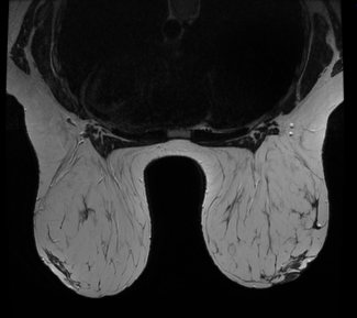
Breast MRI can identify benign conditions like fibroadenomas and cysts, providing detailed characterization that may prevent unnecessary biopsies. It is particularly useful when other imaging results are inconclusive.
Monitoring Treatment Response
In patients undergoing chemotherapy, Breast MRI evaluates treatment efficacy by measuring changes in tumor size and vascularity. It aids in adjusting treatment plans and predicting outcomes.
Benefits of Breast MRI
 Enhanced Imaging Capabilities
Breast MRI offers superior soft-tissue contrast resolution, enabling the detection of small lesions and detailed visualization of breast anatomy. Its multiplanar imaging capability allows for comprehensive assessment from various angles.
Early Detection Advantages
By identifying abnormalities not visible on mammography or ultrasound, Breast MRI reduces the chances of false negatives. Early detection leads to timely intervention, which is crucial for favorable prognoses.
Non-Invasive and Safe
Breast MRI is a non-invasive procedure that does not use ionizing radiation, making it safe for repeated use. The contrast agents used are generally well-tolerated, with minimal risk of allergic reactions.
Personalized Patient Care
The detailed information obtained from Breast MRI enables healthcare providers to tailor treatment plans to the individual patient. It facilitates informed discussions with patients about their options, leading to shared decision-making.
Preparing for a Breast MRI
Patient Preparation Guidelines
Patients are advised to schedule the MRI during the second week of their menstrual cycle to reduce background enhancement. Fasting may be required if sedation is planned. It’s important to inform the provider of any medications, allergies, or kidney issues.
 What to Expect During the Procedure
The procedure typically lasts 30 to 60 minutes. Patients lie face down on a padded table with openings for the breasts, ensuring optimal positioning. Coils are placed around the breasts to enhance signal reception. Earplugs or headphones are provided to mitigate the loud noises produced by the machine.
Post-Procedure Information
After the MRI, patients can usually resume normal activities immediately. If contrast was used, staying hydrated helps eliminate it from the body. Results are typically available within a few days after radiologist review.
Interpretation of Breast MRI Results
Role of the Radiologist
Radiologists specialized in breast imaging interpret the MRI scans. Their expertise is crucial in identifying subtle abnormalities and differentiating benign from malignant lesions. They follow standardized reporting guidelines to ensure consistency.
Understanding the Report
The Breast Imaging Reporting and Data System (BI-RADS) classification provides a standardized assessment:
- BI-RADS 0: Incomplete
- BI-RADS 1: Negative
- BI-RADS 2: Benign findings
- BI-RADS 3: Probably benign
- BI-RADS 4: Suspicious abnormality
- BI-RADS 5: Highly suggestive of malignancy
- BI-RADS 6: Known biopsy-proven malignancy
Communicating Results
Effective communication between radiologists, referring physicians, and patients ensures appropriate follow-up. Providers should discuss findings with patients in a clear and compassionate manner, outlining recommended next steps.
Limitations and Considerations
 False Positives and Negatives
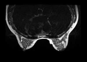
Breast MRI has a high sensitivity but can produce false positives, leading to unnecessary anxiety and procedures. Conversely, false negatives, though less common, can occur. Additional imaging or biopsy may be necessary to confirm findings.
Accessibility and Cost
MRI machines are expensive, and the procedure costs more than other imaging modalities. Insurance coverage varies, potentially limiting patient access. Efforts are ongoing to improve affordability and availability.
Contraindications
Patients with certain implants or devices (e.g., pacemakers) may not be eligible for MRI. Claustrophobia can be a barrier; however, sedation or open MRI machines may be alternatives. Screening for kidney function is important due to the use of contrast agents.
Future Directions in Breast MRI
Technological Advancements
Advances in MRI technology, such as higher-field magnets (7 Tesla scanners) and faster imaging sequences, are enhancing image quality and reducing scan times. These improvements increase patient comfort and diagnostic accuracy.
Research and Clinical Trials
Ongoing research explores new contrast agents and techniques like Magnetic Resonance Spectroscopy (MRS) to provide metabolic information about lesions. Combining MRI with other modalities, like Positron Emission Tomography (PET), is also being investigated.
Impact on Patient Care

Integration of artificial intelligence and predictive analytics holds promise for personalized medicine. These tools may improve risk assessment, detection, and treatment planning, ultimately enhancing patient outcomes.
Breast MRI is a powerful imaging tool offering detailed visualization of breast tissue, crucial for early detection and management of breast diseases. Its benefits include enhanced imaging capabilities and personalized patient care, despite limitations like cost and accessibility.
Collaboration among radiologists, healthcare providers, and patients is essential to maximize the benefits of Breast MRI. Education and open communication foster shared decision-making and improved health outcomes.
Healthcare professionals are encouraged to stay updated with the latest guidelines and technological advancements. Considering Breast MRI in appropriate patient care plans can significantly impact early detection and treatment success.
 Additional Resources
Professional Organizations
- American College of Radiology (ACR): www.acr.org
- Radiological Society of North America (RSNA): www.rsna.org
- Patient Education Materials
- Breast MRI Information for Patients: www.cancer.org
- Understanding Breast MRI: www.breastcancer.org
- Radiology info: https://www.radiologyinfo.org/en/info/breastmr
- References
- American Cancer Society, Breast MRI Scans: https://www.cancer.org/cancer/types/breast-cancer/screening-tests-and-early-detection/breast-mri-scans.html
- National Comprehensive Cancer Network (NCCN). Clinical Practice Guidelines in Oncology: https://www.nccn.org/guidelines/guidelines-process/about-nccn-clinical-practice-guidelines
- European Society of Breast Imaging (EUSOBI). Recommendations for Women’s Imaging: https://www.eusobi.org/news/eusobi-recommendation-breast-cancer-screening-women-with-extremely-dense-breasts/
- Susan G. Koman: https://www.komen.org/breast-cancer/screening/mri/
- National Library of Medicine: https://medlineplus.gov/ency/article/007360.htm
This guide aims to provide a comprehensive overview of Breast MRI, fostering a better understanding among healthcare professionals and patients. For personalized advice and recommendations, consultation with a medical professional is recommended.
Greater Waterbury Imaging Center specializes in MRI scans for patients in the greater Waterbury, CT area. We are committed to providing you with easy access to care, a comfortable exam, and fast, accurate results. Contact GWIC for all your MR imaging needs including breast MRI.

