General Overview of Diseases of the Liver
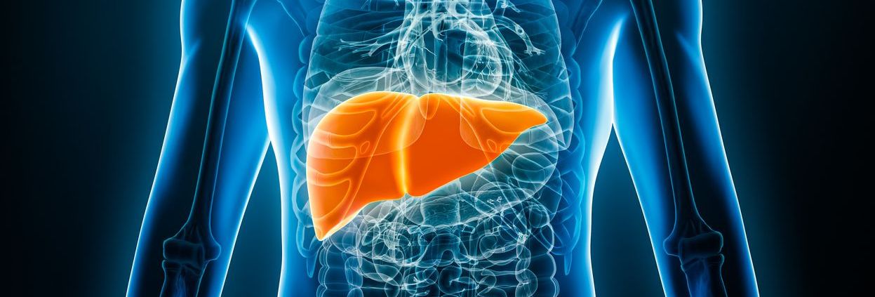
The liver is the largest solid organ in the body, responsible for critical functions including detoxification, the metabolism of fats and carbohydrates, the production of bile, and the synthesis of essential proteins (Ichikawa & Goshima, 2023). Because it filters toxins and processes nutrients, even subtle impairments in liver function can lead to systemic effects. Many liver diseases progress silently, symptoms often appear only once significant damage has occurred, making early detection vital to prevent irreversible injury such as cirrhosis or liver failure (Ichikawa & Goshima, 2023).
Magnetic Resonance Imaging (MRI) offers a non-invasive, highly sensitive method for detecting a wide range of liver pathologies at an early stage, thereby guiding timely interventions and enhancing patient outcomes.
General Description of Diseases of the Liver
1. Fatty Liver Disease (NAFLD/NASH)
Nonalcoholic Fatty Liver Disease (NAFLD) affects an estimated 30% of adults in the United States and up to 25% worldwide, reflecting the global obesity epidemic (Taouli, Ehman, & Reeder, 2009; Younossi, Golabi, & Paik, 2023). NAFLD ranges from simple steatosis (fat accumulation) to Nonalcoholic Steatohepatitis (NASH), in which inflammation and hepatocellular injury accelerate fibrosis. Approximately 20–30% of NASH patients progress to cirrhosis if untreated (Taouli et al., 2009). Early identification of steatosis and fibrosis through MRI enables lifestyle and medical management before advanced disease sets in.
2. Liver Tumors
- Benign lesions, such as hemangiomas, focal nodular hyperplasia, and hepatic adenomas, often require only periodic monitoring once they have been confidently characterized.
- Malignant tumors include Primary Hepatocellular Carcinoma (HCC), the most common primary liver cancer in adults, and metastatic lesions. In 2025, an estimated 42,240 new cases of liver cancer will be diagnosed in the U.S., with HCC comprising about 70% of these (American Cancer Society, 2025). Chronic liver disease (viral hepatitis, NAFLD) remains the principal risk factor for HCC. Accurate MRI-based lesion characterization is crucial for distinguishing between benign and malignant masses and for staging tumor burden.
3. Chronic Liver Diseases (Hepatitis & Cirrhosis)
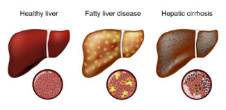
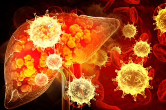
Chronic viral hepatitis (B and C) and long-term alcohol use cause persistent inflammation that may progress over decades to cirrhosis, marked by diffuse fibrosis and regenerative nodules. Cirrhosis elevates risks for portal hypertension, liver failure, and HCC. MRI, including elastography, can noninvasively quantify the fibrosis stage, track disease progression, and reduce the need for some liver biopsies (Taouli et al., 2009).
4. Biliary Tract Abnormalities
Obstructions or strictures in intrahepatic or extrahepatic bile ducts, due to gallstones, cholangiocarcinoma, or postoperative scarring, impair bile flow, leading to jaundice and infection. Magnetic Resonance Cholangiopancreatography (MRCP) provides high-resolution images of the biliary tree without the need for contrast injection, accurately localizing blockages and guiding endoscopic or surgical interventions.
5. Vascular Issues in the Liver
The liver’s dual blood supply via the hepatic artery and portal vein makes it vulnerable to vascular pathologies. Portal vein thrombosis and hepatic artery stenosis can precipitate portal hypertension, variceal bleeding, and ischemic liver injury. Contrast-enhanced Magnetic Resonance Angiography (MRA) delineates vascular anatomy, detects clots or stenoses, and informs interventional radiology planning.
How MRI Is Used to Diagnose and Monitor Diseases of the Liver
Advantages of MRI
- No Ionizing Radiation: Safe for repeated imaging, crucial in chronic disease monitoring, and in younger patients (Ichikawa & Goshima, 2023).
- Superior Soft-Tissue Contrast: Differentiates lesions from normal parenchyma more accurately than CT or ultrasound (Ichikawa & Goshima, 2023).
- Multiparametric Capability: Simultaneously assesses anatomy, tissue composition (fat, iron), perfusion, and mechanical properties (Taouli et al., 2009).
- Operator-Independent Quality: Delivers consistent image quality across centers, reducing variability inherent to ultrasound (Ichikawa & Goshima, 2023).
Key MRI Techniques for Liver Evaluation
- T1- and T2-Weighted Imaging: Baseline anatomical sequences where changes in signal intensity reveal steatosis, edema, or focal lesions.
- Dynamic Contrast-Enhanced MRI: Gadolinium-based agents highlight vascular phases (arterial, portal, delayed), enabling precise lesion characterization (Ichikawa & Goshima, 2023).
- Diffusion-Weighted Imaging (DWI): Detects restricted water movement in malignant tumors and advanced fibrosis; enhances lesion conspicuity without contrast (Taouli et al., 2009).
- Magnetic Resonance Elastography (MRE): Quantifies liver stiffness to stage fibrosis and cirrhosis noninvasively, often replacing biopsy for surveillance (Taouli et al., 2009).
- MR Cholangiopancreatography (MRCP): Heavily T2-weighted sequences that visualize bile and pancreatic ducts fluidly, identifying strictures and stones (Ichikawa & Goshima, 2023).
- Magnetic Resonance Angiography (MRA): High-resolution vascular imaging to assess portal and hepatic arterial flow and patency.
Clinical Applications of Liver MRI
- Diagnosis & Characterization: Differentiates benign from malignant lesions, quantifies fibrosis and steatosis, and evaluates iron overload (Ichikawa & Goshima, 2023).
- Staging and Treatment Planning: Determines tumor size, vascular involvement, and multicentricity in HCC; guides surgical resection, ablation, or transplantation candidacy.
- Monitoring & Follow-Up: Assesses response to antivirals in hepatitis, lifestyle interventions in NAFLD, and locoregional therapies in HCC, adjusting care based on quantitative MRI metrics.
Early detection and precise characterization of liver diseases save lives. Contact Greater Waterbury Imaging Center for world-class MRI services, including MRI of the liver.
References
American Cancer Society. (2025). Cancer Facts & Figures 2025. American Cancer Society
Ichikawa, S., & Goshima, S. (2023). Clinical significance of liver MR imaging. Magnetic Resonance in Medical Sciences, 22(2), 157–175. https://doi.org/10.2463/mrms.rev.2022-0100
Taouli, B., Ehman, R. L., & Reeder, S. B. (2009). Advanced MRI methods for assessment of chronic liver disease. American Journal of Roentgenology, 193(1), 14–27. https://doi.org/10.2214/AJR.09.2601
Younossi, Z. M., Golabi, P., & Paik, J. M. (2023). The global epidemiology of nonalcoholic fatty liver disease (NAFLD) and nonalcoholic steatohepatitis (NASH): A systematic review. Hepatology, 77(4), 1335–1347. https://doi.org/10.1097/HEP.0000000000000004