Table of Contents
Overview of Cardiac MRI
Cardiac Magnetic Resonance Imaging (MRI) is a non-invasive diagnostic tool that provides detailed images of the heart’s structure and function. Utilizing powerful magnetic fields and radio waves, it offers unparalleled clarity without the use of ionizing radiation. Since its introduction, cardiac MRI has become an essential modality in cardiology, aiding in the diagnosis and management of various heart conditions.
Cardiac MRI is used to detect or monitor cardiac disease and to evaluate the heart’s anatomy and function in patients with both heart disease present at birth and heart diseases that develop after birth. Cardiac MRI does not use ionizing radiation to produce images, and it may provide the best images of the heart for certain conditions:
- Evaluating the anatomy and function of the heart chambers, heart valves, size of and blood flow through major vessels, and the surrounding structures such as the pericardium (the sac that surrounds the heart)
- Diagnosing a variety of cardiovascular (heart and/or blood vessel) disorders such as tumors, infections, and inflammatory conditions
- Evaluating the effects of coronary artery disease such as limited blood flow to the heart muscle and scarring within the heart muscle after a heart attack
- Planning a patient’s treatment for cardiovascular disorders
- Monitoring the progression of certain disorders over time
- Evaluating the effects of surgical changes, especially in patients with congenital heart disease
- Evaluating the anatomy of the heart and blood vessels in children and adults with congenital heart disease (heart disease present at birth).
Importance of Cardiac Imaging in Healthcare
Accurate cardiac imaging is crucial for early detection, treatment planning, and monitoring of heart diseases. With heart disease being a leading cause of morbidity and mortality globally, advanced imaging techniques like cardiac MRI play a pivotal role in improving patient outcomes.
Cardiac MRI is a specialized MRI technique focused on capturing high-resolution images of the heart and surrounding vessels. It leverages the magnetic properties of hydrogen atoms in the body’s water and fat molecules, aligning them using a strong magnetic field and then disturbing this alignment with radiofrequency pulses to produce detailed images.
How Cardiac MRI Works
The patient is placed within a large magnet, and when the magnetic field is applied, hydrogen protons align with it. Radiofrequency pulses then alter this alignment, and as the protons return to their original state, they emit signals. These signals are captured and transformed into images by a computer. Adjustments in the technique allow for a specific focus on cardiac tissues, motion, and blood flow.
Modern cardiac MRI machines are equipped with advanced coils and software specifically designed for heart imaging. These machines often have higher magnetic field strengths (1.5 to 3 Tesla) to enhance image quality.
Types of Cardiac MRI Exams
Cardiac MRI encompasses a variety of specialized imaging techniques designed to evaluate different aspects of heart anatomy and function. Each type of exam provides unique information that aids clinicians in diagnosing and managing a wide range of cardiac conditions. Below are the primary types of cardiac MRI exams:
Cine MRI
Cine MRI is akin to creating a movie of the heart in motion. It captures a series of images throughout the cardiac cycle, allowing for detailed visualization of heart function.
- Assessment of Cardiac Function and Motion: Cine MRI is considered the gold standard for evaluating ventricular function. It provides precise measurements of ventricular volumes, ejection fraction, and wall motion. This is crucial for diagnosing conditions such as heart failure, and cardiomyopathies, and assessing the impact of myocardial infarctions.
- Technique: Using balanced steady-state free precession sequences, cine MRI produces high-contrast images between the blood pool and myocardium. Patients are often asked to hold their breath briefly during image acquisition to minimize motion artifacts.
Late Gadolinium Enhancement (LGE)
Late Gadolinium Enhancement is a technique that highlights areas of myocardial scarring or fibrosis by using a contrast agent.
- Detection of Scar Tissue and Fibrosis: After injecting a gadolinium-based contrast agent, damaged myocardial tissues absorb and retain the contrast longer than healthy tissues. This difference in contrast uptake allows for clear visualization of scarred or fibrotic areas within the heart muscle.
- Clinical Applications: LGE is invaluable in diagnosing ischemic heart disease, differentiating between viable and non-viable myocardium, and assessing the extent of damage post-myocardial infarction. It also helps identify non-ischemic cardiomyopathies and infiltrative diseases like amyloidosis.
MRA of the Carotid Arteries
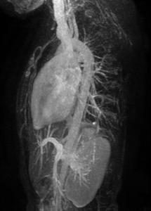
Magnetic Resonance Angiography (MRA) of the Carotid Arteries is a specialized MRI technique focused on visualizing the carotid arteries, which are the major blood vessels supplying oxygen-rich blood to the brain. While it is not a direct component of cardiac MRI, carotid MRA plays a significant role in assessing cardiovascular risk and is often considered in conjunction with cardiac
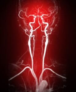
evaluations.
- Non-Invasive Imaging: MRA provides detailed images of the carotid arteries without the need for invasive catheterization or exposure to ionizing radiation.
- Detection of Stenosis and Occlusions: It accurately identifies areas of narrowing or blockages caused by atherosclerotic plaque buildup, which can lead to transient ischemic attacks (TIAs) or strokes.
- Plaque Characterization: Advanced imaging sequences can assess plaque composition, helping to differentiate between stable and unstable plaques that may rupture and cause cerebrovascular events.
T1 and T2 Mapping
T1 and T2 Mapping are quantitative techniques that measure the relaxation times of myocardial tissues, offering insight into tissue composition.
- Quantitative Assessment of Tissue Characteristics:
- T1 Mapping: Measures the T1 relaxation time, which increases in the presence of fibrosis, edema, or infiltration. It is helpful in diagnosing diffuse myocardial diseases like amyloidosis or fibrosis due to hypertensive heart disease.
- T2 Mapping: Sensitive to changes in water content, T2 mapping detects myocardial edema associated with acute injury or inflammation, such as myocarditis or acute myocardial infarction.
These mappings provide a non-invasive means to detect and quantify myocardial abnormalities that may not be apparent with conventional imaging sequences.
4D Flow MRI
4D Flow MRI captures dynamic three-dimensional blood flow over time, providing a detailed hemodynamic assessment. It visualizes complex flow patterns within the heart and vessels, such as vortices and turbulence, making it useful for evaluating valvular diseases, shunts, and aortic pathologies. This technique enhances understanding of flow dynamics, improving diagnosis and treatment planning by offering insights into velocity, direction, and pressure gradients.
Conditions and Diseases Evaluated with Cardiac MRI
Cardiac MRI is a powerful diagnostic tool that offers detailed insights into a variety of heart conditions. Its ability to provide high-resolution images, assess cardiac function, and characterize tissue properties makes it invaluable in diagnosing and managing numerous cardiovascular diseases. Below are the key conditions and diseases effectively evaluated using cardiac MRI:
Coronary Artery Disease
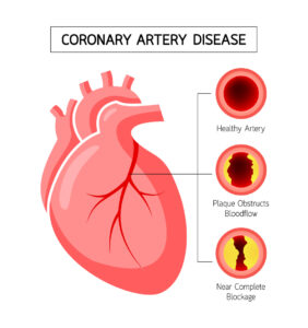
Coronary artery disease (CAD) is characterized by the narrowing or blockage of the coronary arteries due to atherosclerotic plaque buildup.
- Detection of Ischemia and Infarction:
- Ischemia Assessment: Cardiac MRI can detect areas of reduced blood flow (ischemia) during stress perfusion imaging. By comparing rest and stress images, regions with insufficient perfusion indicative of significant coronary artery stenosis can be identified.
- Infarction Characterization: Using Late Gadolinium Enhancement (LGE), cardiac MRI precisely delineates areas of myocardial infarction (heart muscle damage due to lack of blood flow). This helps determine the extent of damage and viability of the myocardium, guiding decisions on revascularization procedures like angioplasty or bypass surgery.
Cardiomyopathies
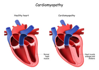
Cardiomyopathies are diseases affecting the heart muscle, leading to altered structure and function.
- Hypertrophic Cardiomyopathy (HCM):
- Assessment of Wall Thickness: Cardiac MRI accurately measures myocardial thickness, identifying areas of hypertrophy that might be missed on echocardiography.
- Detection of Fibrosis: LGE imaging reveals myocardial fibrosis, which is associated with arrhythmias and sudden cardiac death risk.
- Dilated Cardiomyopathy (DCM):
- Evaluation of Chamber Size and Function: Cardiac MRI provides precise measurements of ventricular volumes and ejection fraction, crucial for diagnosing DCM.
- Etiology Differentiation: Helps distinguish between ischemic and non-ischemic causes by assessing tissue characteristics.
- Restrictive Cardiomyopathy:
- Identification of Infiltrative Diseases: Detects conditions like amyloidosis or hemochromatosis through T1 and T2 mapping, which show abnormal tissue properties.
Myocarditis and Pericarditis
Myocarditis is inflammation of the heart muscle, while pericarditis involves inflammation of the pericardial sac surrounding the heart.
- Myocarditis:
- Detection of Inflammation: Cardiac MRI identifies myocardial edema and hyperemia using T2-weighted and early gadolinium enhancement imaging.
- Localization of Injury: LGE can pinpoint areas of necrosis or fibrosis, aiding in differentiating myocarditis from myocardial infarction.
- Clinical Impact: Early diagnosis facilitates appropriate treatment, potentially preventing long-term complications.
- Pericarditis:
- Pericardial Assessment: Evaluates pericardial thickening, effusions, and constrictive physiology.
- Functional Evaluation: Assesses the impact on cardiac filling and function, guiding management decisions.
Congenital Heart Disease
Cardiac MRI is vital for diagnosing and managing congenital heart diseases (CHD), offering:
- Anatomical Detailing: Provides detailed images for diagnosing complex CHD, such as tetralogy of Fallot and ventricular septal defects.
- Functional Assessment: Measures blood flow, shunt volumes, and valve function, and monitors post-surgical outcomes for residual defects or complications.
- Advantages: Non-invasive and safe for repeated use, making it ideal for long-term follow-up, especially in pediatric patients.
Valvular Heart Disease
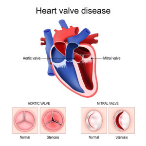
Valvular heart diseases affect blood flow due to valve dysfunction. Cardiac MRI assesses:
- Valve Function: Measures stenosis (narrowing) and quantifies regurgitation (leakage).
- Anatomical Visualization: Provides detailed images of valve structure, identifying issues like prolapse or calcification.
- Cardiac Impact: Evaluates ventricular remodeling and aids in planning surgical or interventional treatments.
Cardiac Tumors and Masses
Cardiac MRI is ideal for evaluating cardiac masses due to superior tissue characterization. It differentiates between benign tumors (like myxomas, fibromas) and malignant ones (such as angiosarcomas), assesses the mass composition (solid, cystic, fatty), and precisely locates it within cardiac structures to evaluate functional impact. Clinically, MRI guides treatment planning (surgical resection or biopsy) and monitors tumor progression or response to therapy.
Vascular Disorders
Cardiac MRI effectively evaluates diseases of the great vessels associated with the heart, including:
- Aortic Aneurysms and Dissections: Provides detailed images of the aortic wall to identify aneurysmal dilatations and intimal flaps, assessing the extent and involvement of branch vessels.
- Pulmonary Hypertension: Evaluates right ventricular size and function; detects enlargement, thrombosis, or other abnormalities in the pulmonary arteries.
- Congenital Vascular Anomalies: Identifies abnormal pulmonary venous return and assesses the severity of aortic coarctation, including collateral circulation.
- Peripheral Vascular Disease (Adjacent to the Heart): Detects obstructions or thrombosis in the superior and inferior vena cava that can affect cardiac function.
Benefits of Cardiac MRI
Cardiac Magnetic Resonance Imaging (MRI) has become an indispensable tool in modern cardiology due to its numerous advantages over other imaging modalities. Its ability to provide detailed images without invasive procedures or radiation exposure makes it highly valuable in diagnosing and managing heart diseases. Below, we delve into the key benefits of cardiac MRI that underscore its significance in cardiac care.
 High-Resolution Imaging
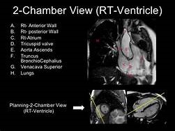
Cardiac MRI stands out for its exceptional image quality, providing superior detail of the heart’s anatomy, including the myocardium, valves, chambers, and surrounding vessels. This high-resolution imaging helps detect subtle abnormalities and differentiates between tissue types, such as fibrosis or edema, which is crucial for diagnosing conditions like myocarditis or cardiomyopathies.
MRI also offers 3D reconstructions, which are essential for assessing complex congenital heart diseases and aiding in surgical planning. This enhanced imaging improves diagnostic accuracy and supports tailored treatment strategies, leading to better patient outcomes.
Non-Invasive and Safe
Cardiac MRI is highly safe and non-invasive, offering significant advantages. Unlike CT scans or nuclear imaging, it uses no ionizing radiation, making it ideal for children and those needing frequent scans. It’s also safer during pregnancy when necessary.
Gadolinium-based contrast agents used in MRI pose a lower risk of allergic reactions than iodine-based ones in CT, with minimal risk of kidney-related side effects. The non-invasive nature of MRI, with no need for catheters or incisions, enhances patient comfort and reduces complications.
Comprehensive Assessment
Cardiac MRI offers a comprehensive evaluation of the heart in a single exam by simultaneously assessing anatomy, function, and tissue properties.
- Structural Assessment: Provides clear images of the heart’s anatomy, identifying defects, wall motion abnormalities, and morphological changes.
- Functional Analysis: Accurately measures ejection fraction, cardiac output, and ventricular volumes.
- Tissue Characterization: Detects fibrosis, scarring, or infiltration, aiding in the diagnosis of cardiomyopathies and myocarditis.
Versatility
Cardiac MRI is highly versatile, making it suitable for a wide range of clinical scenarios.
- Broad Diagnostic Capability: Evaluates various cardiac issues, from congenital heart disease to ischemic conditions, cardiomyopathies, and vascular disorders.
- Customizable Protocols: Imaging sequences can be tailored to assess specific structural, functional, or tissue-related concerns.
Technological Advancements:
- Advanced Imaging: Techniques like T1/T2 mapping, 4D flow imaging, and real-time MRI expand diagnostic options.
- Integration with Other Modalities: Complements data from echocardiography, CT, and nuclear imaging for a comprehensive assessment.
Patient Demographics:
- Pediatric Use: Safe for children, crucial for diagnosing and monitoring congenital heart diseases.
- Alternative for Certain Patients: Ideal for those allergic to CT contrast agents or with limited acoustic windows for echocardiography.
 Reproducibility and Accuracy
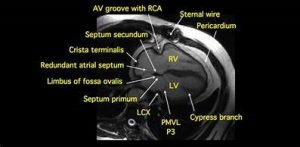
Consistency in cardiac MRI is essential for monitoring disease progression and evaluating treatment effectiveness. It provides highly accurate, reproducible measurements of cardiac dimensions and function, ensuring reliable follow-up across institutions.
This consistency makes MRI ideal for tracking changes in conditions like cardiomyopathies and is valuable in research studies. Clinicians rely on its precision for confident decision-making and early detection of subtle changes, allowing for timely interventions.
Integration with Advanced Technologies
Cardiac MRI is advancing with new technologies like artificial intelligence and machine learning. AI enhances image quality, automates measurements, and improves efficiency, while machine learning models help predict patient outcomes for personalized care. Innovations in functional imaging, such as myocardial strain imaging and molecular imaging, provide deeper insights into heart function and may detect disease at its earliest stages.
Cardiac MRI’s unique combination of safety, detailed imaging, and comprehensive assessment makes it an invaluable tool in the diagnosis and management of heart diseases. Its ability to provide in-depth information without invasive procedures or radiation exposure not only improves patient comfort but also enhances clinical outcomes. Greater Waterbury Imaging Center, GWIC, offers quality cardiac MRI services.

