
Magnetic Resonance Angiography (MRA) at GWIC
What Is Magnetic Resonance Angiography?
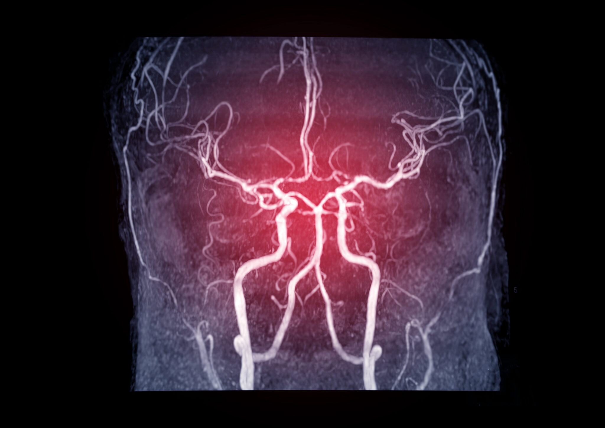
Magnetic resonance angiography, or MRA, is an MRI exam that focuses on the blood vessels. It produces detailed pictures of the arteries, and sometimes the veins, without surgery, without a catheter threaded into an artery, and without the ionizing radiation used in X-ray or CT scans [1]. An MRA can find a narrowed artery, a bulging vessel wall, a blockage, or an abnormal connection between vessels, and it can do all of that from outside the body.
This page is for two readers at once: the patient whose doctor ordered an MRA and wants to know what the exam shows, and the referring clinician who needs to know what Greater Waterbury Imaging Center can image and how. GWIC performs MRA of the brain, neck, chest and aorta, abdomen, and legs on a wide-bore 1.5T scanner in Waterbury, with evening and Saturday hours.
Why Doctors Order an MRA, and Where It Fits
An MRA answers a specific question: is the blood moving through this vessel as it should, and is the vessel itself healthy? Doctors reach for it when they need to see the vessel’s anatomy in detail and want to avoid radiation or an invasive procedure.
Ultrasound is often the first look at a blood vessel, and it’s excellent for the carotid arteries in the neck or vessels near the skin. But ultrasound can’t reach deep vessels well, and it gives a narrower picture than a full cross-sectional scan. MRA shows the whole vascular territory in one exam, including vessels an ultrasound probe can’t reach.
CT angiography is another detailed option, and the two exams overlap. The difference that matters to patients: MRA uses no ionizing radiation and, when contrast is needed, uses gadolinium instead of the iodine-based dye used in a CT scan. Gadolinium is less likely to cause an allergic reaction than iodinated contrast [1]. That makes MRA a strong choice for surveillance, where a patient may be scanned repeatedly over years, and for patients who can’t safely receive iodinated contrast [1]. For aortic aneurysm monitoring, the 2022 ACC/AHA aortic disease guideline lists MRI, which includes MRA, as a reasonable surveillance exam, with imaging repeated every 6 to 24 months when the aorta is stable [2].
Catheter-based angiography still leads in one situation: when the plan is to treat the vessel in the same session, such as placing a stent or coiling an aneurysm. That exam places a catheter directly into the artery. An MRA is a diagnostic imaging exam.
How an MRA Works
An MRA uses the same physics as any MRI. A strong magnetic field aligns the hydrogen atoms in your body. Radio waves briefly knock them out of alignment, and as they settle back, the scanner detects the signals they emit. A computer then turns them into images [1]. The exam adds sequences tuned to blood flow, so the vessels stand out from the surrounding tissue.
Whether an MRA needs contrast depends on which vessels are being imaged. Brain MRA is often performed without contrast, using a technique called time-of-flight that creates images of moving blood on its own [3]. Neck MRA may be performed either way. MRA of the chest, abdomen, and legs is typically performed with gadolinium contrast delivered through a small IV in the arm, with contrast-free options available for patients who need them [3]. If your exam calls for contrast, the technologist places the IV before the scan begins.
The exam itself is easy to go through. You lie on a padded table that slides into the scanner, hold still so the images stay sharp, and you’ll hear the loud knocking the machine makes while it works [1]. You can talk to the technologist the whole time.
Types of MRA by Body Area
MRA is a family of exams grouped by the vessels the scan examines, and each area addresses a different clinical question.
MRA of the Brain
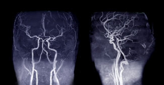
Brain MRA examines the arteries that supply the brain, including the circle of Willis, the ring of vessels at the base of the brain. Doctors order it to screen for an aneurysm, a weak spot in an artery wall that can balloon out, and to identify arteriovenous malformations, which are abnormal tangles connecting arteries and veins [1]. Brain MRA is usually performed without contrast, using time-of-flight imaging [3]. It pairs closely with a standard brain MRI and is covered on GWIC’s brain scan pages when those conditions are the reason for the exam.
MRA of the Carotid Arteries
Carotid MRA images the two large arteries in the neck that carry blood to the brain. Its main job is to detect atherosclerotic plaque that has narrowed the artery, because a tight carotid narrowing is a stroke waiting to happen [1]. When a patient has had a warning stroke or a TIA, carotid imaging tells the doctor whether the narrowing is severe enough to treat. The 2021 AHA/ASA stroke prevention guideline lists MRA as one of the recommended noninvasive ways to screen the carotid arteries after a TIA or anterior-circulation stroke [4].
MRA of the Chest and Aorta
Chest MRA images the aorta, the body’s largest artery, as it leaves the heart and arches down through the chest. It’s used to find and measure an aneurysm, to look for a dissection, which is a tear in the vessel wall, and to follow a known aneurysm over time [1] [2]. Because monitoring requires repeated scans, the radiation-free nature of MRA is a real advantage here. This area typically uses contrast.
MRA of the Abdomen
Abdominal MRA examines the aorta below the diaphragm and the arteries branching from it, including the renal arteries that supply the kidneys. A narrowed renal artery can cause blood pressure that won’t respond to medication, and MRA can show it [1]. When the reason for the scan is the kidneys themselves, that ground is covered on GWIC’s kidney MRI page.
MRA of the Legs
Leg MRA, sometimes called a runoff study, follows the arteries from the pelvis down through the legs to look for narrowing and blockages of peripheral artery disease [1]. It’s the exam that explains why someone’s legs cramp when they walk, and it maps the disease to guide whatever treatment comes next. This area typically uses contrast.
Magnetic Resonance Venography (MRV)
When the question concerns veins rather than arteries, the exam is called magnetic resonance venography, or MRV. Both are magnetic resonance imaging of the vasculature, performed on the same scanner using the same principles. The difference is which side of the circulation is being read. MRV looks for a clot in the deep veins, including those that drain the brain.
Conditions MRA Evaluates
Aneurysms
An aneurysm is a weakened, bulging section of an artery wall that can enlarge and, if it ruptures, cause life-threatening bleeding. MRA shows where an aneurysm is, how large it is, and which vessels it involves, whether in the brain, the aorta, or elsewhere [1]. For aortic aneurysms, MRA is one of the exams used to measure the aorta and track whether it’s growing [2].
Carotid Stenosis and Stroke Risk
Carotid stenosis is a narrowing of the neck arteries caused by plaque buildup, and it’s a major, treatable cause of stroke. MRA measures how severe the narrowing is, which helps a doctor decide whether to treat it and how [1] [4]. When symptoms point to the brain, GWIC’s stroke MRI page explains what the brain scan adds to the vascular study.
Peripheral Artery Disease
Peripheral artery disease narrows the arteries in the legs, causing pain with walking and, when severe, threatening the limb. Leg MRA maps which arteries are narrowed and by how much, so the vascular specialist can plan [1].
Renal Artery Stenosis
A narrowed renal artery can cause high blood pressure that resists medication and can harm kidney function over time. MRA of the abdomen shows the renal arteries and any narrowing [1].
Vascular Malformations and Vasculitis
MRA also helps evaluate malformations of the blood vessels, such as arteriovenous malformations, and vasculitis, which is inflammation of the vessel walls. These conditions have their own dedicated page: GWIC covers them in depth on the malformations of the blood vessels and vasculitis page, where MRA is one of the tools described.
Dissection and Vascular Injury
A dissection is a tear that allows blood to track into the wall of an artery, and trauma can injure vessels in ways that aren’t visible from the outside. MRA can show a dissection flap and assess arteries after an injury [1].
When Timing Matters: Urgent MRA
In a true emergency, such as a stroke in progress or a suspected acute aortic dissection, imaging usually starts with CT angiography because it’s faster to obtain in the moment.
MRA has a specific, urgent role. After a TIA or minor stroke, guidelines call for a prompt diagnostic workup within 48 hours of symptom onset to determine the cause, and noninvasive imaging of the neck arteries with MRA is one of the recommended approaches [4]. MRA is also the preferred option when a patient needs vascular imaging quickly but can’t safely receive the iodinated contrast used in a CT angiogram [1]. GWIC’s STAT MRI service is designed for exactly these time-sensitive orders.
The Exam Experience at GWIC
GWIC performs MRA on a wide-bore 1.5T scanner. The wider opening makes the exam easier for patients who feel claustrophobic, those who are larger, and anyone who has struggled with a narrow-bore machine before. This is why GWIC’s approach to claustrophobia matters as much as the imaging itself. The center offers evening and Saturday hours and sits on the Waterbury Hospital campus with its own gated parking.
Frequently Asked Questions
An MRI images organs and soft tissue. An MRA is a type of MRI aimed specifically at the blood vessels, using sequences tuned to show blood flow [1]. Both run on the same scanner, and a doctor may order them together.
Sometimes. It depends on which vessels are being imaged. Brain MRA is often performed without contrast using a technique called time-of-flight, whereas MRA of the chest, abdomen, and legs usually uses gadolinium contrast administered through an IV in the arm [1] [3]. Your order will specify which.
Both provide detailed images of blood vessels. An MRA uses no ionizing radiation, and when contrast is needed, it uses gadolinium, which is less likely to cause an allergic reaction than the iodine-based dye used in CT angiography [1]. A CT angiogram is often obtained more quickly, which is why emergencies frequently start with it.
Most MRA exams take about 30 to 60 minutes, depending on the area being studied and whether contrast is used. The technologist will provide a time estimate for your specific exam.
Gadolinium contrast requires extra care in patients with reduced kidney function, and in many cases an MRA can be performed without contrast. Tell your doctor and the technologist about any kidney disease before your exam so the appropriate approach is chosen.
Yes. GWIC uses a wide-bore scanner with a wider, more open design, and the team works with patients who feel anxious in enclosed spaces. Our page on MRI and claustrophobia explains the available options.
Schedule Your MRA at Greater Waterbury Imaging Center
If your doctor has ordered an MRA to check for an aneurysm, a narrowed carotid artery, or blocked arteries in your legs, Greater Waterbury Imaging Center performs this exam in Waterbury on a wide-bore 1.5T scanner, with evening and Saturday hours. Call 203-573-7674 to schedule. If you have questions about the exam, claustrophobia, or imaging for a child, contact us, and we’ll walk you through it.
[1] RadiologyInfo.org (RSNA and ACR), “Magnetic Resonance Angiography (MRA).”
https://www.radiologyinfo.org/en/info/angiomr
Supports: Supports the core definition of MRA, the no-radiation and no-catheter points, the gadolinium-versus-iodinated-contrast comparison, MRA as a substitute when iodinated contrast can’t be used, the aneurysm/carotid/AVM/renal/leg indications, and the patient-experience description.
[2] Isselbacher EM, et al. 2022 ACC/AHA Guideline for the Diagnosis and Management of Aortic Disease. Circulation. 2022. (ACC “Ten Points to Remember” summary.)
Supports: Supports MRI/MRA as a reasonable aortic-aneurysm surveillance exam, with imaging every 6 to 24 months when stable.
[3] Bhutani P, et al. “MR Angiogram.” StatPearls (NIH/NCBI Bookshelf).
https://www.ncbi.nlm.nih.gov/books/NBK558984
Supports: Supports the non-contrast-capability of MRA, including time-of-flight technique, and the arterial/venous technique distinctions by territory.
[4] Kleindorfer DO, et al. 2021 Guideline for the Prevention of Stroke in Patients With Stroke and Transient Ischemic Attack. Stroke. 2021 (AHA/ASA).
https://www.ahajournals.org/doi/10.1161/STR.0000000000000375
Supports: Supports MRA as a recommended noninvasive test for carotid stenosis after TIA or anterior-circulation stroke, and the within-48-hours diagnostic-workup timing.