MRI for Diagnosing Alzheimer’s and Dementia
Table of Contents
Dementia is an umbrella term for a decline in cognitive function that interferes with daily life and activities. Among the many types of dementia, Alzheimer’s disease (AD) is the most common, accounting for roughly 60–80% of all dementia cases. According to estimates by the Alzheimer’s Association, millions of Americans—primarily older adults—are currently living with Alzheimer’s disease, and this number is expected to rise as the population ages.
Early detection is crucial because interventions—both pharmacological and non-pharmacological—can potentially slow progression and improve the patient’s quality of life. Magnetic Resonance Imaging (MRI) is often central to this effort; it provides high-resolution images of the brain that help us identify structural changes that may indicate the onset or progression of dementia.
Understanding Dementia and Alzheimer’s
Key Distinctions Among Types of Dementia
Although Alzheimer’s disease is the most frequently diagnosed form of dementia, other types include vascular dementia, Lewy body dementia, and frontotemporal dementia. Each type has characteristic features:
- Alzheimer’s disease: typically presents with memory loss, especially short-term memory, and a gradual progression to language, reasoning, and orientation difficulties.
- Vascular dementia: Often related to strokes or other vascular problems, leading to stepwise declines in cognitive function.
- Lewy body dementia: Marked by fluctuating alertness, visual hallucinations, and Parkinsonian motor symptoms.
- Frontotemporal dementia: This manifests primarily in changes to behavior, personality, and language skills, often at a younger age.
Common Symptoms and Stages
Most dementias share early signs such as forgetfulness, difficulty concentrating, and challenges with complex tasks. Over time, the disease progresses from mild cognitive impairment (MCI)—where individuals can often manage daily routines—to advanced stages, where continuous care and support become necessary.
Risk Factors for Dementia
Several factors increase the likelihood of developing dementia. Advancing age remains the strongest risk factor, but genetics (e.g., APOE4 allele) also plays a role. Lifestyle choices, including diet, exercise, and tobacco use, can influence brain health, while comorbid conditions like hypertension or diabetes may exacerbate cognitive decline.
General Description of Neurological MRI for Dementia and Alzheimer’s
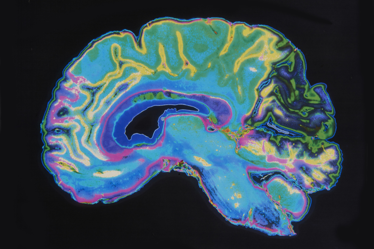
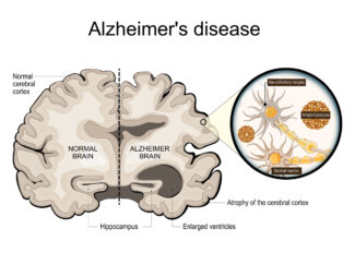
MRI is a preferred imaging modality because it provides detailed views of brain structures without exposing patients to ionizing radiation. In conditions like Alzheimer’s disease, MRI can detect subtle atrophy (shrinkage) in key regions such as the hippocampus, which is essential for forming new memories. These structural changes can precede clinical symptoms, making MRI pivotal for early detection.
Key MRI Sequences and Techniques
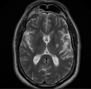 Typical MRI protocols for dementia evaluation include T1-weighted, T2-weighted, and FLAIR sequences to visualize brain tissue and identify lesions or atrophy. Volumetric MRI adds quantitative measures of brain regions like the hippocampus. Advanced imaging techniques—such as diffusion tensor imaging (DTI)—further highlight white matter integrity, revealing microstructural changes that may correlate with cognitive deficits.
Patient Experience and Safety
Patients undergoing an MRI lie comfortably on a motorized table that slides into the scanner’s cylindrical magnet. Although some individuals experience claustrophobia, the procedure is safe and painless. For those patients who experience claustrophobia, GWIC offers Non-Claustrophobic MRI, with wider openings for a more comfortable MRI experience.
While MRI does not use harmful radiation, patients should inform the technologist of any metal implants or devices to ensure safety.
How MRI Is Used to Diagnose and Monitor Dementia and Alzheimer’s
Diagnosing Dementia and Alzheimer’s
MRI findings can help confirm or rule out certain conditions when a patient exhibits memory or behavioral concerns. A radiologist or neurologist reviews images for hallmark patterns of brain atrophy, particularly in the medial temporal lobe. This diagnostic information is combined with clinical assessments and neuropsychological evaluations to arrive at a more definitive diagnosis.
Monitoring Disease Progression
MRI is also valuable for tracking changes in brain structure over time. For instance, periodic scans can measure the rate of atrophy or the development of additional abnormalities. These objective markers guide treatment decisions and support clinical judgment about medication adjustments, therapy, or lifestyle interventions.
Correlation with Other Diagnostic Tools
Other imaging studies, such as positron emission tomography (PET), can detect amyloid or tau protein deposition, both of which are strongly linked to Alzheimer’s disease. Laboratory tests (blood or cerebrospinal fluid biomarkers) and comprehensive cognitive assessments further clarify the diagnosis and help tailor treatment plans.
Future Directions in Imaging
Ongoing research aims to refine imaging techniques, including functional MRI (fMRI) and higher-field strength MRI scanners, which can reveal earlier or subtler changes. These advancements may allow for more precise characterizations of disease progression and a greater ability to personalize therapies.
Additional Considerations for Patients and Caregivers
Preparation for an MRI Examination 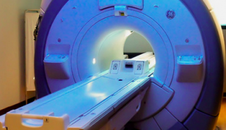
Most patients need only remove metal objects and wear comfortable clothing. If a contrast agent is required, you may be asked about allergies or kidney function in advance. Please follow any instructions provided by your healthcare team to ensure a smooth and accurate imaging session.
Importance of Follow-Up
Given the progressive nature of dementia, regular follow-up with your neurologist or primary care physician is essential. Consistent monitoring—through clinical evaluations and periodic imaging—offers the best chance to adjust treatments promptly and maintain the highest possible quality of life.
Support and Resources
Local and national Alzheimer’s or Dementia advocacy organizations offer resources for caregivers and patients. These groups provide education, support networks, and practical tips for daily living. Counseling services, caregiver support groups, and nutritional guidance can also play vital roles in patient well-being.
Conclusion and Next Steps
The use of MRI in diagnosing and monitoring Alzheimer’s disease and dementia cannot be overstated. By revealing structural brain changes even before significant symptoms emerge, MRI empowers clinicians and patients to make well-informed decisions about treatment and lifestyle modifications.
If you suspect cognitive impairment or have concerns about memory decline, it is important to seek medical evaluation early. Speak to your healthcare provider to schedule an MRI or consultation and take advantage of the latest advances in neurological imaging to preserve and optimize cognitive health. Contact GWIC for all your MR imaging needs.

