Table of Contents
MRI for Brain Infections
Brain infections, or central nervous system infections, occur when pathogens such as bacteria, viruses, fungi, or parasites invade the brain tissue or surrounding structures. These infections can manifest in various ways, with conditions including meningitis, encephalitis, and brain abscesses.
Types of Brain Infections:
- Bacterial infections: Examples include bacterial meningitis and brain abscesses.
- Viral infections: Such as herpes encephalitis, West Nile virus, and HIV-related encephalopathy.
- Fungal infections: Cryptococcal meningitis and aspergillosis are common in immunocompromised individuals.
- Parasitic infections: Conditions such as neurocysticercosis and toxoplasmosis.
Causes and Risk Factors: Brain infections can result from various pathogens entering through the bloodstream, direct injury, or spreading from nearby infected structures (such as sinuses or ears). Immunocompromised patients, elderly individuals, and infants have increased susceptibility.
Clinical Symptoms and Presentation: Symptoms often include headache, fever, seizures, altered mental status, confusion, and neurological deficits. Timely diagnosis and treatment are critical to minimizing permanent neurological damage or death.
Neurological MRI for Brain Infections
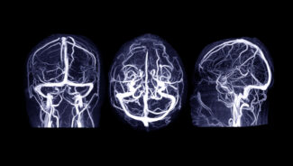
Magnetic Resonance Imaging (MRI) plays a crucial role in diagnosing and managing brain infections due to its superior capability to visualize soft tissue and detect subtle changes in brain structure and composition.
Role of MRI in Brain Infection Evaluation: MRI is highly sensitive, allowing early detection of infections before extensive damage occurs. Unlike CT scans, MRI provides detailed images without radiation exposure and better identifies inflammation, edema, and subtle brain abnormalities.
MRI Techniques Used for Brain Infections:
- T1 and T2-weighted imaging: Basic imaging to identify anatomical abnormalities.
- Fluid-Attenuated Inversion Recovery (FLAIR): Highlights inflammation and edema clearly.
- Diffusion-weighted imaging (DWI): Particularly valuable in identifying acute brain abscesses and areas of restricted diffusion, indicative of infection.
- Gadolinium-enhanced MRI: Contrast enhancement identifies breakdown of the blood-brain barrier, common in infections.
- MR Spectroscopy (MRS): Provides biochemical information that helps differentiate infection from other neurological conditions.
Typical MRI Appearance of Brain Infections:
- Bacterial infections: Characteristic findings include ring-enhancing lesions indicative of abscess formation.
- Viral infections: Typically involve specific regions, such as temporal lobes in herpes encephalitis, often with swelling and enhanced contrast uptake.
- Fungal infections: May show multiple small enhancing lesions, especially in immunocompromised patients.
- Parasitic infections: Often present as cystic lesions or calcifications, as seen in neurocysticercosis.
How MRI is Used to Diagnose and Monitor Brain Infections
MRI serves as an essential diagnostic and monitoring tool in the management of brain infections.
Initial Diagnosis: MRI is often the initial imaging modality when a brain infection is suspected due to its ability to quickly identify and differentiate infections from other conditions like stroke or tumors.
Assessing Extent and Severity: MRI accurately visualizes the location, size, and extent of brain infections, evaluates complications such as edema, hydrocephalus, or infarction, and guides intervention plans.
Guiding Treatment Decisions: Imaging results inform decisions regarding medication (antibiotics, antivirals, antifungals, antiparasitics) or surgical interventions (such as abscess drainage or biopsy).
Monitoring Treatment Response: Serial MRI examinations monitor the efficacy of treatments, track progression or regression of infections, and guide modifications in therapy, enhancing patient outcomes.
Specific Brain Infections and MRI Findings
- Bacterial Infections:
- Meningitis: MRI typically demonstrates leptomeningeal enhancement.
- Brain Abscess: Distinctive ring-enhancing lesions with restricted diffusion on DWI.
- Viral Infections:
- Herpes Encephalitis: MRI shows characteristic involvement of the temporal lobes, frequently bilateral, with inflammation and swelling.
- HIV Encephalopathy: Diffuse white matter changes indicating progressive neurological impairment.
- Fungal Infections:
- Cryptococcosis: Characteristically seen as basal ganglia lesions or enhancing nodules.
- Parasitic Infections:
- Neurocysticercosis: Cystic lesions and calcifications often have a “starry sky” MRI appearance.
- Toxoplasmosis: Typically multiple ring-enhancing lesions in basal ganglia or gray-white matter junctions.
Advantages of MRI for Brain Infection Management
- High sensitivity and specificity compared to other imaging modalities.
- Non-invasive, painless, and without exposure to ionizing radiation.
- Superior delineation of anatomical and pathological details.
Patient Preparation and MRI Examination Expectations
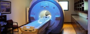 Patients should wear comfortable clothing without metal objects. MRI exams typically last between 30-60 minutes, and sedation is available for those experiencing anxiety or difficulty staying still. Contrast material may be administered intravenously to enhance imaging details, which is generally safe and well-tolerated.
Neurological MRI at Greater Waterbury Imaging Center
Greater Waterbury Imaging Center is dedicated to providing expert care through advanced MRI technology including neurological MRI, ensuring accurate diagnosis and effective management of brain infections. Our experienced radiologists collaborate closely with referring physicians, delivering individual patient care that prioritizes accuracy, comfort, and positive outcomes.
Contact us for all your MR imaging needs including neurological MRI for brain infections.