MRI for Diagnosing and Monitoring Stroke
Table of Contents
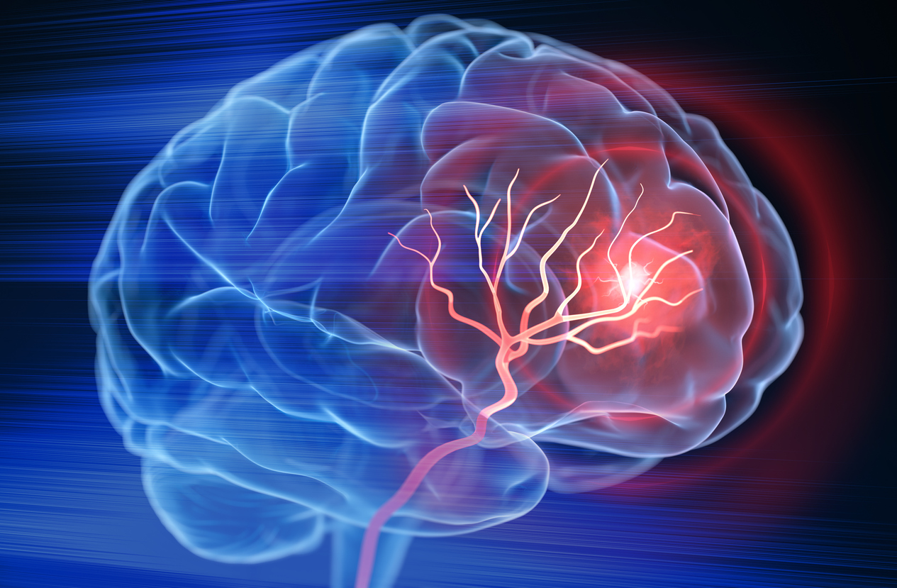
A stroke occurs when blood flow to part of the brain is disrupted, leading to a loss of oxygen and nutrients in the affected area. This disruption can lead to permanent brain damage if not addressed promptly. Strokes are typically classified into two main categories:
- Ischemic (caused by a blood clot or blockage)
- Hemorrhagic (caused by a ruptured blood vessel)
Recognizing stroke symptoms quickly and seeking immediate medical attention can significantly improve outcomes. Medical imaging, particularly Magnetic Resonance Imaging (MRI), plays an essential role in detecting the precise nature and location of a stroke, which is vital to guide effective treatment and recovery strategies.
Understanding Stroke
Types of Stroke
- Ischemic Stroke: This is the most common type, occurring when a clot blocks an artery, cutting off blood supply. Early intervention with medications such as tissue plasminogen activator (tPA) can help dissolve the clot if administered quickly.
- Hemorrhagic Stroke: These strokes happen when a weakened blood vessel in the brain ruptures, causing bleeding into or around the brain tissue. Risk factors often include hypertension and aneurysms.
- Transient Ischemic Attack (TIA): Commonly referred to as a “mini-stroke,” a TIA involves a temporary blockage with symptoms that resolve, usually within minutes to hours. Even though TIAs do not typically result in permanent damage, they serve as a warning sign and warrant further medical evaluation.
Signs and Symptoms
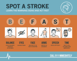
Patients often experience sudden onset of weakness or numbness (especially on one side of the body), confusion or difficulty speaking, vision changes, dizziness, or severe headache. A helpful tool for recognizing stroke symptoms is the BE FAST acronym:
Balance, Eyes, Face drooping, Arm weakness, Speech difficulties, and Time to call emergency services.
Early intervention remains the key to preventing long-term complications and preserving brain function. Learn more about risk factors, finding support, and preventing a second stroke.
Risk Factors
Stroke risk factors are categorized into modifiable and non-modifiable factors.
Modifiable Risk Factors:
-
Hypertension (High Blood Pressure): The leading cause of stroke; managing blood pressure is crucial.
-
Diabetes: Increases the risk of stroke due to its impact on blood vessels.
-
High Cholesterol: Contributes to plaque buildup in arteries, leading to blockages.
-
Smoking: Damages blood vessels and raises blood pressure.
-
Obesity and Physical Inactivity: Linked to other risk factors like hypertension and diabetes.
-
Excessive Alcohol Consumption: Can increase blood pressure and the risk of stroke.
Non-Modifiable Risk Factors:
-
Age: Risk increases with age, particularly after 55.
-
Family History and Genetics: A family history of stroke can increase risk.
-
Gender: Women may have unique risk factors, including pregnancy-related conditions and hormonal therapies.
Hypertension (high blood pressure) is the leading preventable risk factor for stroke. Other modifiable contributors include diabetes, high cholesterol, smoking, and obesity. Family history and genetic predispositions also play a role, highlighting the importance of regular check-ups and a healthy lifestyle. Understanding and managing these risk factors is essential for stroke prevention. Learn more about Stroke Awareness.
Neurological MRI for Stroke
Why MRI Is Used
MRI offers detailed images of brain structures without exposing patients to ionizing radiation. In the setting of stroke, MRI can identify subtle changes—sometimes within minutes of onset—making it highly valuable in acute situations. Rapid and accurate identification of ischemic versus hemorrhagic stroke ensures that patients receive the most appropriate therapy.
Key MRI Sequences and Techniques
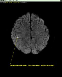
- Diffusion-Weighted Imaging (DWI): Detects early ischemic changes by highlighting areas where water molecule movement is restricted, often indicating acute stroke.
- Perfusion MRI: Assesses blood flow to determine if there is salvageable brain tissue (the “penumbra”) around the core of the stroke.
- T2/FLAIR: Useful for identifying edema and chronic lesions.
- MR Angiography (MRA): Provides a detailed view of blood vessels, pinpointing arterial blockages or aneurysms.
Patient Comfort and Safety
During an MRI, the patient lies on a table that slides into a cylindrical magnet. Although some individuals are concerned about confined spaces, the procedure is painless, and sedation options are available if needed. Patients should inform the technologist of any implants, pacemakers, or other devices for safety reasons.
How MRI Is Used to Diagnose and Monitor Various Types of Stroke
 Diagnosing Ischemic vs. Hemorrhagic Stroke
Differentiating between a clot-based blockage and a blood vessel rupture is critical to determining treatment. MRI sequences such as DWI and FLAIR can help confirm ischemic stroke, while other sequences or MRI-based angiographic views can highlight bleeding or vascular malformations.
Monitoring and Follow-Up Scans
Patients who have suffered a stroke may require additional MRI examinations to track their recovery and identify potential complications, such as swelling or further bleeding. These follow-up images also aid in adjusting rehabilitation plans and medication regimens.
Correlation with Other Imaging Techniques
Computed Tomography (CT) scans often remain the first-line imaging modality in emergency settings due to speed and widespread availability. However, once a patient is stable, MRI frequently provides more detailed insight. Carotid artery ultrasound might also be used alongside MRI to detect plaque or other vessel abnormalities in the neck.
Future Directions and Emerging Technologies
Researchers are exploring faster, more advanced MRI sequences and leveraging artificial intelligence to interpret diffusion and perfusion images. These innovations hold promise for more precise diagnosis and individualized treatment options in acute stroke care.
Additional Considerations for Patients and Caregivers
Preparation for an MRI Examination (Brief)
In most cases, patients only need to remove metallic objects and inform the imaging team of any implants or medical devices. Those who experience anxiety or claustrophobia may receive mild sedation, ensuring a smooth and accurate examination. Learn more about how to prepare for your MRI exam.
Importance of Rehabilitation and Ongoing Care
Post-stroke rehabilitation often involves physical, occupational, and speech therapies to regain function and independence. Lifestyle modifications—such as controlling blood pressure, quitting smoking, and maintaining a healthy diet—reduce the risk of a second stroke. Caregivers and patients can benefit from local support groups and online resources that provide education and emotional support.
 Neurological MRI
MRI is a powerful tool in the diagnosis, management, and follow-up of stroke. By rapidly identifying the type of stroke, MRI assists physicians in determining the most effective treatment plan, ultimately improving patient outcomes. If you or a loved one experiences any signs of stroke, seek immediate medical attention.
Recognized for Excellence in Stroke Care
 Waterbury Hospital has once again received the American Heart Association’s Get With The Guidelines® – Stroke award, recognizing our continued commitment to delivering high-quality, evidence-based stroke care. This national recognition honors hospitals that consistently follow the latest research-based clinical guidelines to improve outcomes for stroke patients.
The Get With The Guidelines® program is a hospital-based initiative developed by the American Heart Association and American Stroke Association to promote adherence to proven treatment protocols. Facilities that earn this designation demonstrate excellence in areas such as rapid stroke assessment, timely administration of clot-busting treatments like tPA, and comprehensive discharge planning for recovery and rehabilitation.
This achievement reflects our ongoing dedication to improving survival rates, reducing disability, and ensuring the best possible care for every stroke patient. It reinforces our role as a trusted provider for advanced stroke evaluation, including neurological MRI imaging for stroke.
GWIC offers neurological MRI services, including scans of the brain, inner auditory canal, orbits, head, neck, and spine. Please speak with your healthcare provider to discuss concerns about stroke risk. Contact us to schedule all your MR imaging needs, including neurological MRI for stroke.