High Resolution MRI to Evaluate the Brain Vasculature
MRI or Magnetic Resonance Imaging is an excellent diagnostic tool to evaluate the vasculature in the brain. MRI uses a strong magnetic field and RF or radio frequency waves which the signals are processed to provide vasculature images in the brain. This tool is used to evaluate many diseases of the vasculature including intracranial artery disease. The use of magnetic resonance imaging and magnetic resonance angiography produces highly detailed images of the cardiovascular system including the vasculature. And unlike x-rays, MRI doesn’t use any radioactive rays.
Magnetic Resonance Imaging for Intracranial Artery Disease
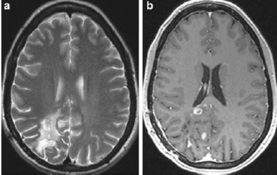
At Greater Waterbury Imaging Center, we use an MRI technique to evaluate intracranial artery disease specifically for differentiating vessel wall patterns of vasculitis of the CNS (Central Nervous System) and cerebral vasoconstriction syndrome. Using our 1.5T high-resolution MR imaging system, our imaging protocol consists of T1-weighted sequences using contrast agents and with fat suppression techniques, and magnetic resonance angiography (see more information on MRA) of the circle of Willis. Characteristics of the vessel walls which can be evaluated with this technique include enhancement, wall thickening, and lumen narrowing.
This technique allows our radiologists to visualize the wall and lumen of large intracranial vessels and can potentially increase the specificity of imaging to diagnose disparate vascular diseases with similar angiographic findings. If you have patients who need their vasculature evaluated for vessel wall changes, please contact Greater Waterbury Imaging Center to discuss how we can help in evaluating patients with intracranial artery disease.