
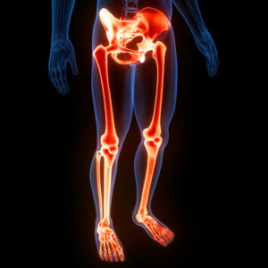
MRI is particularly valuable for evaluating the lower extremities, including the hips, knees, ankles, femur, tibia, fibula, and feet. This advanced imaging technique provides high-resolution images of bones, muscles, tendons, ligaments, and other soft tissues, making it an essential tool for diagnosing and managing many orthopedic conditions.
Imaging such as MRI lower extremity exams help diagnose and treat various conditions affecting the following areas of the body. Learn more about the many conditions affecting the lower parts of the body and how magnetic resonance imaging can help diagnose, treat, and manage these conditions.
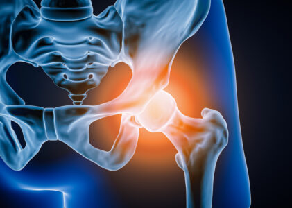
Hip MRI
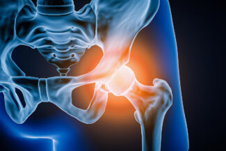
A Hip MRI lower extremity exam is a specialized imaging test used to assess various conditions affecting the hip joint and surrounding structures. This type of MRI is crucial for diagnosing the underlying causes of hip pain, evaluating bone fractures, identifying degenerative joint disorders, detecting inflammation, diagnosing infections, and uncovering tumors. Below is a detailed description of each indication and the utility of MRI in diagnosing and evaluating these conditions.
Pain
Hip or Buttock Pain, Hip-related Groin Pain, or Pain After Hip Surgery:
Hip MRI is highly effective in identifying the root causes of hip or buttock pain and hip-related groin pain. The detailed images can reveal soft tissue abnormalities, labral tears, cartilage damage, and other structural issues that might not be evident through physical examination or other imaging techniques. Additionally, MRI is crucial for post-surgical assessment to ensure proper healing and to identify any complications such as infections or improper placement of surgical implants.
Bone Fractures
Fractures Not Visible on X-rays or Other Imaging Tests:
Hip MRI is instrumental in detecting bone fractures that are not visible on X-rays or other imaging modalities. Stress fractures and occult fractures, which are common in athletes and elderly patients, can be precisely identified using MRI. This allows for early and accurate diagnosis, facilitating appropriate and timely treatment to prevent further complications.
Degenerative Joint Disorders
Arthritis or Other Joint Abnormalities:
MRI is a key tool in evaluating degenerative joint disorders, such as osteoarthritis. It provides a comprehensive view of the joint, revealing the extent of cartilage loss, bone marrow lesions, synovial inflammation, and other joint abnormalities. This detailed assessment aids in determining the severity of the condition and in planning effective treatment strategies, including conservative management and surgical interventions.
Inflammation
Bursitis or Tendonitis:
Hip MRI is valuable for diagnosing inflammatory conditions such as bursitis and tendonitis. MRI of the lower extremities can detect inflammation of the bursae (fluid-filled sacs that cushion the bones, tendons, and muscles near joints) and tendons, providing clear images of swelling, fluid accumulation, and tissue damage. This information is crucial for devising appropriate treatment plans to reduce inflammation and alleviate pain.
Infections
Osteomyelitis and Other Infections:
A lower extremity MRI is highly sensitive in detecting infections in the hip joint and surrounding areas. Conditions like osteomyelitis (infection of the bone) can be accurately diagnosed with MRI, which shows detailed images of bone marrow and soft tissue changes associated with infection. Early detection through MRI allows for prompt treatment with antibiotics or surgical intervention, preventing the spread of infection and serious complications.
Tumors
Primary Tumors and Metastases:
Hip MRI is essential for identifying primary tumors and metastatic lesions in the hip region. It provides high-resolution images that help in differentiating benign from malignant tumors, assessing the size and extent of the tumor, and planning biopsy or surgical procedures. MRI’s ability to detect soft tissue involvement and bone marrow infiltration makes it a critical tool in the comprehensive evaluation of suspected tumors.
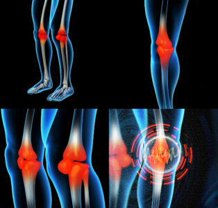
Knee MRI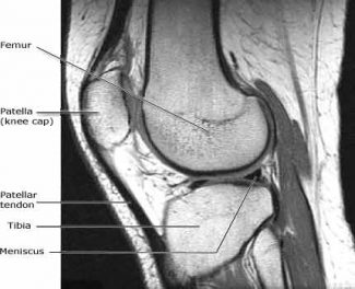
A Knee MRI is a specialized MRI lower extremity imaging test that provides detailed images of the knee joint and surrounding structures. This non-invasive procedure is crucial for diagnosing and evaluating a variety of knee-related conditions, offering clear insights into issues that might not be detectable through physical examination or other imaging modalities. Below, we explore the specific indications for a Knee MRI and the utility of MRI in diagnosing and managing these conditions.
Knee Pain
Unexplained Chronic Pain, or Acute Pain Preventing Walking, or Causing Redness, Swelling, or Deformity:
Knee MRI is particularly useful for patients experiencing chronic knee pain with no clear diagnosis from other imaging tests or physical examinations. It helps in identifying underlying causes such as cartilage damage, meniscal tears, and soft tissue inflammation. For acute pain that is severe enough to prevent walking or is accompanied by redness, swelling, or deformity, MRI provides a detailed assessment to identify fractures, ligament tears, or other traumatic injuries that require immediate attention.
Knee Instability
The Knee Giving Out for No Reason, or Suspected Internal Derangement:
Patients who experience knee instability, where the knee gives out unexpectedly, can benefit significantly from a Knee MRI. This imaging technique is adept at detecting internal derangements, including ligament tears (such as ACL, PCL, MCL, or LCL tears), meniscal tears, and other structural abnormalities that contribute to instability. Accurate diagnosis through MRI aids in planning appropriate surgical or non-surgical treatments to restore knee stability.
Soft Tissue Damage
Damage to Ligaments, Cartilage, Tendons, or Meniscus:
MRI is the gold standard for assessing soft tissue damage in the knee. It provides high-resolution images of the ligaments, cartilage, tendons, and menisci, allowing for precise identification of tears, strains, and degenerative changes. This is particularly important for athletes and active individuals, as it guides the treatment plan, whether it involves physical therapy, surgical repair, or other interventions to restore knee function and prevent further injury.
Bone Injuries
Fractures Not Visible on X-rays, or Osteochondral Injuries:
For bone injuries that are not clearly visible on X-rays, such as stress fractures or occult fractures, Knee MRI is an essential diagnostic tool. It can also detect osteochondral injuries, where both the bone and the cartilage are damaged. These injuries often require detailed imaging for proper diagnosis and treatment planning, ensuring that patients receive appropriate care to promote healing and prevent complications.
Other Conditions
Infections, Tumors, Arthritis, Fluid Buildup, or Sports Injuries Like Sprains:
Knee MRI is invaluable in diagnosing a range of other conditions affecting the knee joint. In cases of suspected infections, such as septic arthritis or osteomyelitis, MRI provides detailed images that reveal the extent of infection and inflammation. It is also critical in identifying tumors, distinguishing between benign and malignant growths, and assessing their impact on surrounding tissues.
For arthritis, MRI lower extremity imaging offers a comprehensive view of joint degeneration, helping to evaluate the severity of the condition and guide treatment options. Fluid buildup in the knee, known as effusion, can be assessed with MRI to determine its cause and guide appropriate interventions. Sports injuries, such as sprains, can be accurately diagnosed with MRI, revealing the extent of ligament damage and other related injuries, facilitating effective treatment and rehabilitation plans.
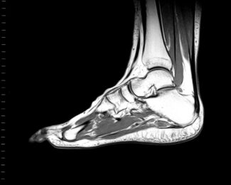
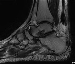 Ankle MRI
An Ankle MRI is a specialized imaging test that provides comprehensive and detailed images of the ankle joint and surrounding structures. This non-invasive MRI lower extremity procedure is pivotal for diagnosing and evaluating various ankle-related conditions, offering clarity that might not be achievable through other imaging modalities or physical examinations. Below, we detail the specific indications for an MRI of the ankle and the utility of MRI in diagnosing and managing these conditions.
Tendon Disorders
Partial and Complete Tears, Tendinitis, Tendinopathy, Tenosynovitis, Subluxation, and Dislocation in the Achilles, Posterior Tibial, and Peroneal Tendons:
Ankle MRI is highly effective in diagnosing tendon disorders, including partial and complete tears, tendinitis, tendinopathy, tenosynovitis, subluxation, and dislocation. The detailed images provided by MRI can reveal the extent of tendon damage in the Achilles, posterior tibial, and peroneal tendons, which are critical for ankle stability and function. This precise assessment helps in planning appropriate treatments, whether it be conservative management or surgical intervention, to restore tendon integrity and function.
Bone Abnormalities
Osteomyelitis, Occult Fractures, and Stress Fractures:
MRI is a crucial tool for identifying bone abnormalities in the ankle that might not be visible on X-rays or other imaging tests. It is particularly useful for diagnosing osteomyelitis (infection of the bone), occult fractures, and stress fractures. Early detection of these conditions through MRI allows for timely treatment, preventing further complications and promoting effective healing.
Soft-Tissue Abnormalities
Plantar Fasciitis, Plantar Fibromatosis, Interdigital Neuromas, and Tarsal Tunnel Syndrome:
An MRI lower extremity exam of the ankle provides high-resolution images of the soft tissues, making it invaluable for diagnosing conditions such as plantar fasciitis, plantar fibromatosis, interdigital neuromas, and tarsal tunnel syndrome. These conditions can cause significant pain and dysfunction, and accurate imaging is essential for identifying the underlying issues. MRI aids in developing targeted treatment plans, whether through physical therapy, injections, or surgical interventions, to alleviate symptoms and restore normal function.
Other Injuries
Ligament Injuries, Sprains, Instability, Ankle Impingement, and Suspected Chondral Injuries:
MRI lower extremity is the preferred imaging method for assessing ligament injuries, sprains, and instability in the ankle. It can detect tears, strains, and other injuries to the ligaments that might not be evident on physical examination alone. Additionally, MRI is effective in diagnosing ankle impingement, where soft tissues are pinched within the joint, and suspected chondral injuries, involve damage to the cartilage surfaces. Accurate diagnosis through MRI facilitates appropriate treatment planning, helping to restore stability and function to the ankle joint.
Pain and Swelling
Unexplained Pain or Swelling in the Ankle or Rearfoot:
For patients experiencing unexplained pain or swelling in the ankle or rearfoot, MRI is a crucial diagnostic tool. It provides a comprehensive view of the ankle’s internal structures, identifying potential causes such as inflammation, soft tissue injuries, or bone abnormalities. This detailed imaging helps in developing an effective treatment plan to address the underlying cause of pain or swelling and improve patient outcomes.
Femur MRI
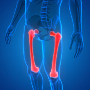
A Femur MRI is a specialized MRI lower extremity test that offers detailed images of the femur (thigh bone) and surrounding structures. This non-invasive procedure is essential for diagnosing and evaluating a variety of conditions affecting the femur, providing insights that might not be achievable through other imaging techniques or physical examinations. Below, we explore the specific indications for a Femur MRI and the utility of MRI in diagnosing and managing these conditions.
Bone Abnormalities
Fractures, Stress Fractures, Avascular Necrosis, and Marrow Edema Syndromes:
Femur MRI is highly effective in diagnosing bone abnormalities such as fractures, stress fractures, avascular necrosis (AVN), and marrow edema syndromes. While X-rays and CT scans can detect obvious fractures, MRI is superior in identifying subtle stress fractures and early-stage AVN, which might not be visible on other imaging modalities. MRI can also detect marrow edema syndromes, which involve the accumulation of excess fluid in the bone marrow. Early and accurate diagnosis of these conditions through MRI allows for timely and appropriate treatment, preventing further complications and promoting optimal healing.
Soft Tissue Abnormalities
Tumors, Abscesses, Ulcers, and Infections:
A lower extremity MRI is invaluable for identifying soft tissue abnormalities in the femur, such as tumors, abscesses, ulcers, and infections. It provides high-resolution images that help differentiate between benign and malignant tumors, assess the size and extent of abscesses, and detect the presence of ulcers and infections. This detailed imaging is critical for developing effective treatment plans, whether surgical or non-surgical, to address these conditions and improve patient outcomes.
Muscle and Tendon Abnormalities
Strains, Tears, Tendonitis, and Tendonopathy:
Femur MRI is a key tool for assessing muscle and tendon abnormalities, including strains, tears, tendonitis, and tendinopathy. It offers detailed images of the muscles and tendons, revealing the extent of injury and inflammation. This information is crucial for devising appropriate treatment strategies, which may include physical therapy, medication, or surgical intervention, to restore muscle and tendon function and prevent further injury.
Ligament Abnormalities
Tears and Injuries:
MRI is the preferred imaging method for diagnosing ligament abnormalities in the femur. It can detect tears and injuries to the ligaments that might not be evident through physical examination or other imaging techniques. Accurate diagnosis of ligament injuries through MRI is essential for planning effective treatment, whether conservative or surgical, to restore stability and function to the affected area.
Degenerative Disorders
Arthritis:
MRI is a valuable tool for evaluating degenerative disorders such as arthritis. It provides a comprehensive view of the joint, showing the extent of cartilage loss, bone marrow lesions, synovial inflammation, and other joint abnormalities. This detailed assessment helps in determining the severity of the condition and in planning effective treatment strategies, including conservative management and surgical interventions, to alleviate symptoms and improve joint function.
Congenital and Developmental Conditions
Birth Defects:
Femur MRI is essential for diagnosing congenital and developmental conditions affecting the femur. It provides detailed images that help identify birth defects and other developmental abnormalities, allowing for early and accurate diagnosis. This information is critical for planning appropriate interventions and treatments to address these conditions and improve patient outcomes.
Tibia and Fibula MRI
A Tibia and Fibula MRI is a specialized imaging test that provides detailed images of the tibia (shin bone) and fibula, along with the surrounding tissues. This non-invasive procedure is crucial for diagnosing and evaluating various conditions affecting these bones and their associated structures. Below, we explore the specific indications for a Tibia and Fibula MRI and the utility of MRI lower extremity in diagnosing and managing these conditions.
Bone Problems
Bone Contusions, Osteonecrosis, Marrow Edema Syndromes, Stress Fractures, Osteoarthritis, and Tumors:
Tibia and Fibula MRI is highly effective in diagnosing a range of bone problems. It can detect bone contusions (bruises), which are often missed on X-rays and other imaging modalities. MRI is also superior in identifying osteonecrosis (bone death due to reduced blood flow) and marrow edema syndromes, which involve fluid accumulation in the bone marrow. Stress fractures, which are small cracks in the bone that can occur from overuse or repetitive activity, are clearly visible on MRI, allowing for early diagnosis and treatment.
For patients with osteoarthritis, lower extremity MRI provides a detailed view of the joint, showing the extent of cartilage loss, bone changes, and synovial inflammation. This helps in determining the severity of the condition and planning effective treatment strategies. MRI is also invaluable in detecting tumors in the tibia and fibula, differentiating between benign and malignant growths, and assessing their impact on surrounding tissues.
Soft Tissue Issues
Sprains, Strains, Tears, Nerve Compression, and Tendonitis:
MRI is the preferred imaging method for assessing soft tissue issues in the tibia and fibula region. It provides high-resolution images of the muscles, tendons, ligaments, and nerves, allowing for precise identification of sprains, strains, tears, and nerve compression. Tendonitis, which is inflammation of the tendons, can also be accurately diagnosed with MRI. This detailed imaging is crucial for developing targeted treatment plans to address these issues, whether through physical therapy, medication, or surgical intervention.
Other Conditions
Infections, Ligament Tears, Cysts, and Birth Defects:
Tibia and Fibula MRI are invaluable for diagnosing other conditions such as infections, ligament tears, cysts, and birth defects. In cases of suspected infections, MRI provides detailed images that reveal the extent of infection and inflammation in the bones and surrounding tissues. This allows for prompt treatment with antibiotics or surgical intervention to prevent the spread of infection and serious complications.
Ligament tears in the tibia and fibula region can be precisely identified with MRI, facilitating appropriate treatment planning to restore stability and function. MRI is also effective in detecting cysts, which are fluid-filled sacs that can develop in the bone or soft tissue, and assessing their impact on surrounding structures.
For congenital and developmental conditions, MRI lower extremity offers detailed imaging that helps identify birth defects and other abnormalities, allowing for early and accurate diagnosis. This information is critical for planning appropriate interventions and treatments to address these conditions and improve patient outcomes.
Foot MRI
A Foot MRI is an MRI lower extremity specialized imaging test that provides detailed images of the foot’s bones, tendons, and surrounding soft tissues. This non-invasive procedure is essential for diagnosing and evaluating various conditions affecting the foot, offering insights that might not be achievable through other imaging techniques or physical examinations. Below, we explore the specific indications for a Foot MRI and the utility of MRI in diagnosing and managing these conditions.
Bone and Tendon Abnormalities
Osteomyelitis, Occult Fractures, and Tears in the Achilles, Tibialis Posterior, and Peroneal Tendons:
Foot MRI is highly effective in diagnosing bone and tendon abnormalities. It is particularly useful for detecting osteomyelitis (infection of the bone) and occult fractures, which may not be visible on X-rays or other imaging modalities. MRI provides detailed images of the bones, allowing for early and accurate diagnosis and facilitating timely treatment to prevent further complications.
Tendon abnormalities, such as tears in the Achilles, tibialis posterior, and peroneal tendons, are also clearly visible on MRI. This imaging technique can reveal the extent of tendon damage, helping in the planning of appropriate treatment strategies, whether conservative management or surgical intervention, to restore tendon function and prevent further injury.
Soft Tissue Abnormalities
Plantar Fasciitis, Plantar Fibromatosis, Interdigital Neuromas, and Tarsal Tunnel Syndrome:
MRI is invaluable for diagnosing soft tissue abnormalities in the foot. Conditions such as plantar fasciitis and plantar fibromatosis, which involve inflammation and fibrous tissue growth in the plantar fascia, can be accurately identified with MRI. This detailed imaging helps in developing targeted treatment plans to alleviate symptoms and restore normal function.
Interdigital neuromas, benign growths of nerve tissue between the toes, and tarsal tunnel syndrome, a condition caused by compression of the tibial nerve, are also effectively diagnosed with MRI lower extremity. This imaging technique provides high-resolution images of the soft tissues, allowing for precise identification of these conditions and guiding appropriate treatment interventions.
Other Conditions
Muscle Tears, Infections, Tumors, Ligament and Tendon Damage, Stress Fractures, Damaged Cartilage, Sports-Related Injuries, Extent of Arthritis, Infection or Fluid Build-Up, and Persistent Pain After Surgery:
Foot MRI is essential for diagnosing a wide range of other conditions affecting the foot. It is highly effective in identifying muscle tears, infections, and tumors, providing detailed images that reveal the extent of the abnormality and its impact on surrounding tissues. This information is crucial for developing effective treatment plans to address these conditions and improve patient outcomes.
For ligament and tendon damage, MRI offers detailed imaging that can identify tears, strains, and other injuries that might not be evident through physical examination alone. This is particularly important for athletes and active individuals, as it guides the treatment plan, whether it involves physical therapy, surgical repair, or other interventions, to restore function and prevent further injury.
Stress fractures, small cracks in the bone that occur from overuse or repetitive activity, are clearly visible on MRI. This allows for early diagnosis and treatment to prevent further damage and promote healing. Damaged cartilage, which can occur from injury or degenerative conditions, can also be accurately assessed with MRI, facilitating appropriate treatment planning.
For sports-related injuries, MRI is the preferred imaging method for evaluating the extent of damage to the foot’s structures. It provides detailed images that guide the treatment and rehabilitation process, helping athletes return to their activities as quickly and safely as possible.
MRI is also invaluable in assessing the extent of arthritis, revealing the severity of joint degeneration, and helping to guide treatment options. Additionally, it effectively detects infection or fluid build-up in the foot, allowing for prompt and appropriate treatment to prevent further complications.
Finally, for patients experiencing persistent pain after surgery, MRI lower extremity exams can provide detailed images that reveal any underlying issues, such as improper healing, infection, or hardware problems, that may be causing the pain. This information is crucial for developing effective treatment plans to address these issues and alleviate the pain.
This is a detailed overview of the many types of orthopedic MRI exams for the lower extremities. Each section gives critical information about how MRI technology helps in diagnosing and managing complex conditions, helping to provide proper care for patients. Contact GWIC for all your orthopedic imaging needs including MRI lower extremity exams.