Table of Contents
- 1 Orthopedic MRI of the Spine: Advanced Imaging to Diagnose and Evaluate Many Conditions
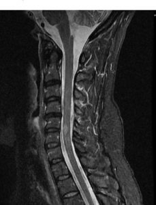
- 2 MRI of the Cervical Spine
- 2.1 1. Evaluation of Pain, Numbness, Tingling, or Weakness
- 2.2 2. Assessment of Chronic Diseases of the Nervous System
- 2.3 3. Diagnosis of Tumors or Cancer in the Spine
- 2.4 4. Detection of Infections in the Vertebrae or Surrounding Tissues
- 2.5 5. Identification of Structural Abnormalities and Degenerative Conditions
- 3 MRI of the Thoracic Spine
- 3.1 1. Detection of Fractures
- 3.2 2. Identification of Infections
- 3.3 3. Assessment of Swelling
- 3.4 4. Evaluation of Tumors
- 3.5 5. Detection of Unusual Curves in the Spine
- 3.6 6. Identification of Spinal Stenosis
- 3.7 7. Diagnosis of Arthritis
- 3.8 8. Evaluation of Nerve Damage
- 3.9 9. Assessment of Spinal Disc Problems
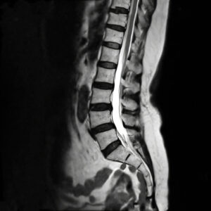
- 4 MRI of the Lumbar Spine
Orthopedic MRI of the Spine: Advanced Imaging to Diagnose and Evaluate Many Conditions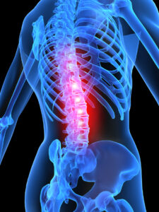
Magnetic Resonance Imaging (MRI) is a powerful diagnostic tool that uses magnetic fields and radio waves to create detailed images of the body’s internal structures. An orthopedic MRI of the spine is invaluable in diagnosing and evaluating a wide range of conditions and diseases that affect the spinal column and its surrounding tissues. This page provides an overview of how MRI is used in orthopedic evaluations of the spine, with a focus on the three major sections: the Cervical Spine, Thoracic Spine, and Lumbar Spine.
MRI of the Cervical Spine
The cervical spine, which comprises the upper part of the spine in the neck region, plays a crucial role in supporting the head and enabling a range of movements. Given its significance, issues in the cervical spine can lead to a variety of symptoms and require accurate diagnosis through advanced imaging techniques like MRI.
An MRI of the cervical spine can help to diagnose and assess chronic conditions to further direct treatment whether physical therapy, medications, or surgical intervention.
1. Evaluation of Pain, Numbness, Tingling, or Weakness
One of the most common reasons for ordering an MRI of the cervical spine is to evaluate persistent or unexplained symptoms such as pain, numbness, tingling, or weakness in the neck, shoulders, or arms. These symptoms often indicate nerve compression or damage, which can be caused by conditions such as herniated discs or spinal stenosis. An MRI provides a detailed view of the spinal cord, nerve roots, and intervertebral discs, helping physicians pinpoint the exact cause of these symptoms.
 2. Assessment of Chronic Diseases of the Nervous System
Chronic diseases such as multiple sclerosis (MS) can affect the nervous system, including the spinal cord. An MRI of the cervical spine is crucial in diagnosing and monitoring MS and other neurological conditions. The MRI can detect lesions, inflammation, or other abnormalities in the spinal cord, which are critical for determining the progression of the disease and guiding treatment decisions.
3. Diagnosis of Tumors or Cancer in the Spine
MRI is the gold standard for identifying tumors or cancerous growths in the spine. The detailed images produced by an MRI of the spine can reveal the size, location, and nature of tumors within the cervical spine, whether they originate in the vertebrae, spinal cord, or surrounding tissues. Early detection through MRI is vital for effective treatment planning, which may include surgery, radiation, or chemotherapy.
4. Detection of Infections in the Vertebrae or Surrounding Tissues
Infections such as spinal epidural abscesses can be life-threatening if not diagnosed promptly. An MRI of the cervical spine can detect these infections by showing inflammation, abscesses, or other signs of infection in the vertebrae or surrounding soft tissues. This allows for timely intervention, which is critical in preventing complications such as spinal cord compression or sepsis.
5. Identification of Structural Abnormalities and Degenerative Conditions
The cervical spine is susceptible to a variety of structural abnormalities and degenerative conditions, which can cause significant discomfort and impair mobility. MRI of the spine is used to diagnose conditions such as:
- Spinal Stenosis: A narrowing of the spinal canal that can compress the spinal cord or nerve roots, leading to pain and neurological deficits.
- Herniated Discs: Displacement of the intervertebral disc material can irritate nearby nerves, causing pain and neurological symptoms.
- Arthritis: Degeneration of the joints in the cervical spine can lead to pain, stiffness, and reduced range of motion.
- Spondylolisthesis: A condition where one vertebra slips forward over the one below it, which can cause pain and spinal instability.
- Spondylosis: Degenerative changes in the spine, particularly in the intervertebral discs and facet joints, leading to chronic pain and stiffness.
Each of these conditions can be accurately diagnosed with MRI, providing a clear image of the spine’s anatomy and the extent of any degenerative or structural changes. This information is essential for developing an appropriate treatment plan, whether it involves physical therapy, medication, or surgical intervention.
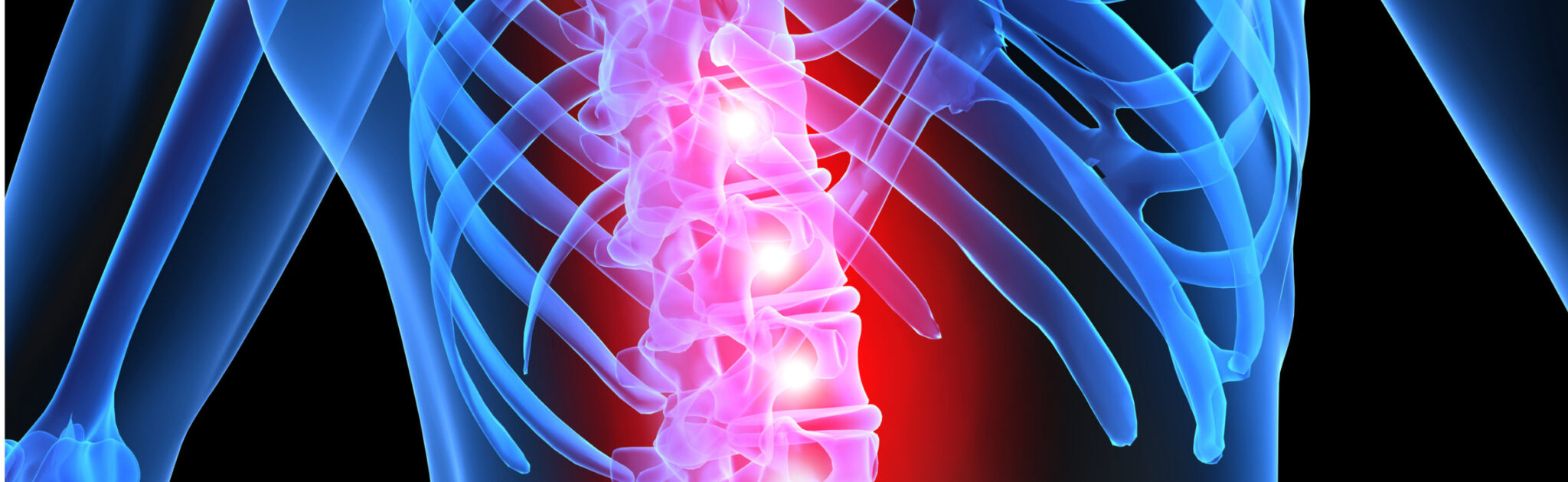
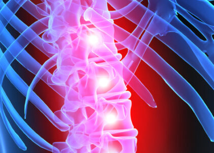
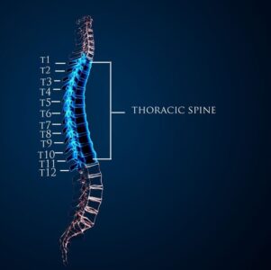 MRI of the Thoracic Spine
The thoracic spine, located in the middle of the back, is a complex structure that supports the upper body and protects the spinal cord. While it is less mobile than the cervical or lumbar regions, the thoracic spine can still be affected by various conditions that require precise imaging for diagnosis and treatment planning.
An MRI of the thoracic spine identifies a variety of issues such as fractures, infection, swelling, tumors, structural abnormalities, arthritis, nerve damage, and more.
1. Detection of Fractures
Fractures in the thoracic spine can result from trauma, osteoporosis, or other underlying conditions. MRI is often employed to evaluate the extent of these fractures, particularly when they are not clearly visible on X-rays. MRI can provide detailed images that help determine the severity of the fracture, the involvement of surrounding soft tissues, and whether there is any compression of the spinal cord or nerve roots.
2. Identification of Infections
Infections in the thoracic spine, such as vertebral osteomyelitis or spinal epidural abscesses, can have serious consequences if not diagnosed and treated promptly. An MRI is highly effective in detecting infections, as it can show signs of inflammation, abscess formation, and other indicators of infection in both the vertebrae and the surrounding soft tissues. Early detection through MRI is crucial for preventing complications and guiding appropriate treatment.
3. Assessment of Swelling
Swelling within the thoracic spine can be indicative of various conditions, including trauma, infection, or inflammatory diseases. MRI of the spine can visualize soft tissue swelling and its effects on the spinal cord and nerves, helping to determine the underlying cause and the most appropriate course of treatment.
4. Evaluation of Tumors
Tumors in the thoracic spine, whether benign or malignant, require accurate diagnosis and evaluation to determine the appropriate treatment plan. MRI provides detailed images that can reveal the size, location, and characteristics of tumors within the spine, spinal cord, or surrounding tissues. This imaging is essential for planning surgical intervention, radiation therapy, or other treatments.
5. Detection of Unusual Curves in the Spine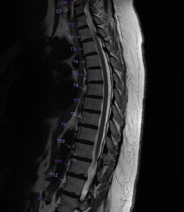
Conditions such as scoliosis or kyphosis, which involve abnormal curvature of the spine, can significantly impact a person’s posture, mobility, and overall health. MRI is used to assess the degree and nature of these curvatures, as well as to evaluate any associated spinal cord or nerve involvement. This information is vital for determining the best course of treatment, which may include bracing, physical therapy, or surgery.
6. Identification of Spinal Stenosis
Spinal stenosis, a narrowing of the spinal canal, can occur in the thoracic spine and lead to compression of the spinal cord or nerve roots. MRI of the spine is the preferred imaging modality for diagnosing spinal stenosis, as it provides detailed images of the spinal canal, allowing physicians to assess the severity of the condition and plan for treatment, which may range from conservative management to surgical intervention.
7. Diagnosis of Arthritis
Arthritis can affect the joints of the thoracic spine, leading to pain, stiffness, and reduced mobility. MRI can detect the early signs of arthritis, such as joint space narrowing, bone spurs, and inflammation. By visualizing the extent of arthritic changes, MRI aids in tailoring treatment plans that may include medication, physical therapy, or in severe cases, surgery.
8. Evaluation of Nerve Damage
Nerve damage in the thoracic spine can result from various conditions, including trauma, compression, or degenerative diseases. MRI of the spine is an essential tool for assessing nerve damage, as it can visualize the nerves, spinal cord, and surrounding structures in detail. This helps physicians understand the extent of nerve involvement and plan for appropriate interventions, whether surgical or non-surgical.
9. Assessment of Spinal Disc Problems
The spinal discs in the thoracic region, like those in other parts of the spine, can be subject to conditions such as bulging, slipped, or ruptured discs. These issues can cause significant pain and neurological symptoms. MRI is the most effective imaging technique for evaluating spinal discs, as it provides clear images of the disc structure, including any herniation, degeneration, or compression of nearby nerves. Accurate diagnosis of disc problems is essential for developing a treatment plan that may include physical therapy, pain management, or surgery.
MRI of the Lumbar Spine
The lumbar spine, located in the lower back, is a common site of pain and discomfort due to its role in supporting much of the body’s weight and enabling movement. An MRI of the lumbar spine is often used to diagnose the underlying causes of symptoms and to plan appropriate treatment strategies.
1. Assessment of Back Pain or Leg Pain, Weakness, or Numbness
Lower back pain and radiating leg pain, often accompanied by weakness or numbness, are frequent complaints that lead to the use of MRI in evaluating the lumbar spine. These symptoms can result from a variety of conditions, including nerve compression, herniated discs, or degenerative diseases. MRI provides detailed images that can help identify the precise cause of pain, allowing for targeted treatment such as physical therapy, medication, or surgery.
2. Diagnosis of Herniated Discs, Tumors, or Infections
MRI is highly effective in detecting and evaluating common issues such as herniated discs, tumors, or infections within the lumbar spine. A herniated disc, which occurs when the soft inner material of a spinal disc bulges out, can press on nearby nerves, causing pain and neurological symptoms. Tumors, whether benign or malignant, and infections in the lumbar region can also cause significant problems and require a prompt diagnosis. MRI offers the detailed imaging necessary to identify these conditions and assess their severity.
3. Evaluation of Bladder and Bowel Control Issues
Problems with bladder and bowel control can sometimes be linked to issues in the lumbar spine, such as nerve compression or spinal cord abnormalities. MRI is crucial in evaluating these symptoms as it can reveal conditions like cauda equina syndrome, a serious disorder that requires immediate medical attention. By providing clear images of the spinal cord and surrounding structures, MRI helps in diagnosing the underlying cause of these symptoms and planning the appropriate treatment.
4. Detection of Fractures
Fractures in the lumbar spine can occur due to trauma, osteoporosis, or other conditions. MRI is used to assess these fractures, especially when they are not clearly visible on other imaging modalities like X-rays. MRI can show the extent of the fracture, any involvement of the spinal cord or nerve roots, and whether there is any associated soft tissue damage. This information is essential for guiding treatment, which may range from conservative management to surgical intervention.
5. Identification of Spinal Stenosis
Spinal stenosis, characterized by a narrowing of the spinal canal, can occur in the lumbar spine and lead to symptoms such as back pain, leg pain, and difficulty walking. MRI is the preferred method for diagnosing spinal stenosis because it provides detailed images of the spinal canal, allowing physicians to see the extent of the narrowing and any associated nerve compression. This information is crucial for determining the best treatment approach, whether it involves physical therapy, pain management, or surgery.
This is a detailed overview of the various types of orthopedic MRI of the spine, including cervical, thoracic, and lumbar. Each section gives critical information about how MRI technology helps in diagnosing and managing complex conditions of the spine. Contact GWIC for all your orthopedic imaging needs including cervical, thoracic, or lumbar orthopedic MRI of the spine.