

MR Enterography
MR Imaging of the Small Bowel
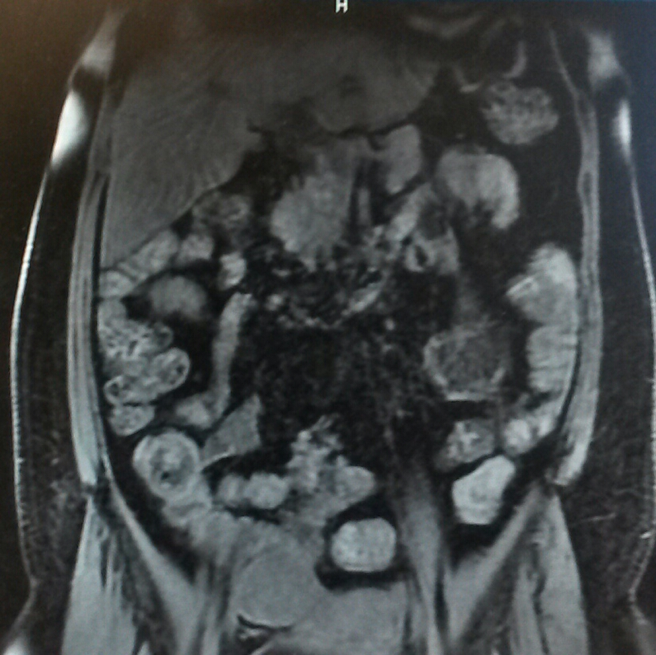 Cross-sectional imaging techniques of suspected small-bowel disorders allows for the visualization of the entire bowel without the overlapping loops of the bowel. Currently, there are two recognized MR imaging techniques used to provide the visualization and they are differentiated by the way the contrast material is administered either orally or through a nasogastric tube. In healthcare, it is always important and a goal to reduce the patient’s exposure to ionizing radiation if possible and this can be accomplished by using imaging methods such as Magnetic Resonance. MR is an imaging modality with the not only the advantage of no ionizing radiation but the ability to visualize the small bowel with cross-sectional information.
Advantages and Limitations of Using MRI for Imaging the Small Bowel
As mentioned, the significant advantages of MR imaging include the lack of ionizing radiation and the superior tissue contrast provided by the technology. In many small-bowel disease processes, the symptoms and manifestations of the disease appear at an early age. This would necessitate the frequent monitoring and assessment of the progression of the small-bowel disease for complications and its response to therapy.
Compared to CT (computed tomography), MR imaging provides superior tissue contrast and the safety associated with MR intravenous contrast does in general make MR imaging of the small bowel feasible in patients where they cannot tolerate the CT contrast agents or it’s contraindicated. Another major advantage is that many abnormalities may be detected with MR without having to use any contrast material if contraindicated for that patient. Specifically, pregnant patients or those at risk for developing nephrogenic disorders, would benefit from small bowel assessment without contrast using MR.
In summary, MR Enterography is an imaging study that is usually done with intravenous contrast but when contrast is not an option for the patient, the study can be done effectively by imaging the patient after the large consumption of water by the patient.