MRI Bilateral Breasts with and without IV Contrast
HISTORY: BILATERAL INFLAMMATORY BREAST CANCER
TECHNIQUE: Multiplanar images of the breasts were obtained at 1.5 Tesla on a dedicated breast coil prior to and following administration of 10 Ml OF Gadavist intravenous contrast. MRI Bilateral Breasts with and without IV Contrast.
COMPARISON: Mammogram 9/11/2017
FINDINGS:
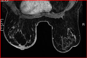
There are a few foci of metal artifact at the 12:00 position of the left breast, 6 cm from the nipple, with corresponding parenchymal architectural distortion consistent with postsurgical scarring. There are additional foci of metal artifact along the left axillary tail and left axilla consistent with surgical clips. The area of parenchymal scarring at the 12:00 position of the left breast measures approximately 2.2 x 1.6 cm. There is increased vascularity within the scar. There is extensive diffuse left breast skin thickening with increased vascularity throughout the skin which may represent inflammation or inflammatory carcinoma. There is also increased left nipple enhancement which may also be secondary to inflammation or carcinoma. There is diffuse left breast edema. The left breast parenchyma is diffusely nodular. The nodular areas within all four quadrants of the left breast demonstrate increased enhancement, although on the time-activity curve, there is increased delayed enhancement which is less characteristic for malignancy, although is nonspecific.
There is moderate right breast skin thickening and mild skin enhancement. There is right breast edema. There is an ovoid area of increased parenchymal enhancement at the 10:00 position of the right breast measuring 4.5 x 2.2 cm, and located approximately 5 to 8 cm from the nipple. There is an additional 1.0 cm area of nodular enhancement posteriorly in the right breast at the 10:00 position, approximately 12 cm from the nipple. Both of these nodular areas of enhancement in the right breast demonstrate heterogeneous enhancement. There are several enlarged right axillary lymph nodes. The largest of these measure 1.9 cm in short axis diameter.
This study was evaluated using a computer aided detection software program.
IMPRESSION: Bilateral skin thickening and increased enhancement suspicious for inflammatory carcinoma. There is additional left breast nipple enhancement. The breasts are edematous. There is enhancing scar at the 12:00 position of the left breast. There is right axillary lymphadenopathy. There are nodular enhancing areas in both breasts which are nonspecific although are suspicious for multifocal malignancy. The largest of these areas of enhancement is seen at the 10:00 position of the right breast measuring up to 4.5 x 2.2 cm.
ACR BIRADS: #5: Highly suggestive of malignancy.