

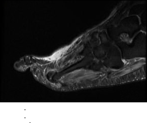
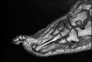
MRI of the Right Foot
HISTORY: Osteomyelitis
TECHNIQUE: Multiplanar images of the right foot were obtained at 1.5 Tesla without IV contrast.
BONES: There is a bone marrow edema throughout the right first distal phalanx. No focal cortical erosion or replacement of the normal fatty marrow is seen. There is no fracture or subluxation. There are moderate midfoot degenerative changes.
TENDONS: The flexor and extensor tendons are intact.
LIGAMENTS: There is no evidence of ligament rupture.
SOFT TISSUES: There is dorsal soft tissue edema within the distal foot and edema within the first digit. There is no evidence of a discrete abscess.
IMPRESSION: Bone marrow edema of the right first distal phalanx which is nonspecific and may represent early osteomyelitis, however there is no associated cortical erosion or infiltration of the normal fatty marrow.