

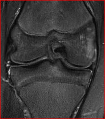
MRI Image of a Right Knee Contusion
Age: 13 yrs
History: Right Knee Internal Derangement, Twisting Injury, Heard a ‘pop’.
Comparison: None
Technique: Multiplanar images of the right knee were obtained at 1.5 Tesla without IV contrast.
FINDINGS:
 Ligaments:
ACL: The anterior cruciate ligament is intact.
PCL: The posterior cruciate ligament is intact.
MCL: The medial collateral ligament is intact.
LCL: The lateral collateral ligament is normal.
MINISCI:
Medial Meniscus: The medial meniscus in intact.
Lateral Meniscus: The lateral meniscus is intact.
BONES: There is a small area of bone marrow edema, along the most medial aspect of the distal femur, involving the medial femoral epiphysis consistent with an area of bone bruising. No significant arthropathy is seen.
SOFT TISSUES: The quadriceps and patellar tendons are intact. No soft tissue abnormality is seen. There is no significant joint effusion.
IMRESSION: Small area of bone marrow edema, along the most medial aspect of the distal femur, involving the medial femoral epiphysis, consistent with an area of bone bruising. No evidence of ligamentous or meniscal injury.
For more information on MRI imaging services at Greater Waterbury Imaging Center, visit our clinical section of the website.