
HISTORY: PALPITATIONS

COMPARISON: None.
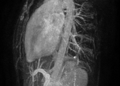
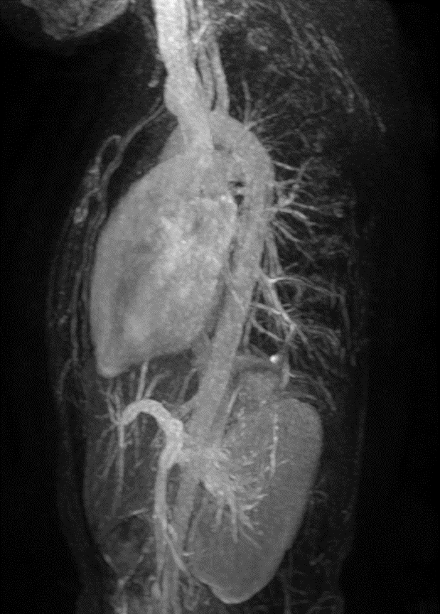
TECHNIQUE: Multiplanar images of the heart were obtained at 1.5 Tesla. Morphologic and dynamic cine imaging was performed in multiple projections. 10 mL of intravenous Gadavist was administered for delayed enhancement imaging. Flow quantification sequences were obtained. Post processing included flow and volume calculations, which were performed by the interpreting physicians on an independent workstation.
FINDINGS:
Thoracic Aorta: The thoracic aorta is normal in caliber. No aneurysm or dissection is seen. The ascending aorta measures 2.9 cm in diameter.
Pulmonary artery: The main pulmonary artery is normal, measuring 2.5 cm in diameter. Systemic and pulmonary venous return: Conventional. Interatrial septum: Intact.
Cardiac chambers: Normal atrioventricular and ventriculo-arterial concordance.
Coronary arteries: Normal origins.
Left ventricle: The left ventricular size is mildly enlarged. Left ventricular systolic function is normal. No segmental wall motion abnormalities are seen. There is normal myocardial wall thickness throughout the left ventricle. T2-weighted imaging demonstrates no high signal intensity to suggest the presence of myocardial edema. Post contrast images only demonstrat
e normal physiologic delayed myocardial enhancement at the right ventricular insertion site anteriorly. No thrombus is visible in the left ventricle. There is no systolic anterior motion of the mitral valve or outflow tract obstruction.
LV MEASUREMENTS:
BSA: 1.47 sq. m., weight: 104 lb, height: 63 in.
LVEDV: 129 ml. LVEDVI: 88 ml/sq.m. (Normal < 78; mildly increased: 78-100; moderately increased: 101-120; severely increased > 120 ml/sq.m)
LVESV: 58 ml. LVESVI: 40 ml/sq.m.
LVSV: 71 ml. LVSVI: 49 ml/sq.m.
LVEF: 55% (Normal >54%; mildly depressed: 40-54%; moderately depressed: 30-39%; severely depressed <30%)
EDD: 47 mm (normal: 40-52 mm)
ESD: 32 mm (normal: 23-35 mm)
Anterior septal wall: 8 mm (normal<12 mm)
Posterior septal wall: 7 mm (normal<11 mm)
LV mass: 66 g.
LV mass index: 45 g/sq.m. (Normal < 60; mildly increased: 60-74; moderately increased: 75-100; severely increased > 100 g/sq.m)
Cardiac output: 6.0 L/min
Cardiac index: 4.07 L/min/sq.m.
Right ventricle: The right ventricular size is normal. Right ventricular systolic function is globally normal.No segmental wall motion abnormalities or aneurysms are seen. Post-contrast images demonstrate no delayed myocardial enhancement. No thrombus is visible in the right ventricle.
RV MEASUREMENTS:
RVEDV: 127 ml. RVEDVI: 86 ml/sq.m. (Normal: 47-103; mildly increased: 104-153; moderately increased: 154-206; severely increased > 206 ml/sq.m)
RVESV: 64 ml. RVESVI: 43 ml/sq.m.
RVSV: 63 ml. RVSVI: 43 ml/sq.m.
RVEF: 50% (Normal >46%; mildly depressed: 36-46%; moderately depressed: 25-35%; severely depressed <25%)
Left atrium: The left atrial size is normal, measuring 2.8 cm AP in end systole (Normal: 2.8-4.0). No thrombus is visible in the left atrium.
Right atrium: The right atrial size is normal, measuring 3.0 cm AP in end systole on the four-chamber view (Normal: 2.8-4.5). No thrombus is visible in the right atrium.
Aortic valve: There is no significant aortic stenosis or regurgitation. Normal-appearing tricuspid aortic valve. Phase-contrast imaging at the ascending aorta reveals a forward flow of 75 cc/heart beat.
Pulmonic valve: There is no significant pulmonic stenosis or regurgitation. Phase-contrast imaging at the main pulmonary artery reveals a forward flow of 81 cc/heart beat.
Qp/Qs: Normal.
Mitral valve: There is no significant mitral stenosis or regurgitation.
Tricuspid valve: There is no significant tricuspid stenosis or regurgitation.
Pericardium: No pericardial thickening, enhancement or pericardial effusion is identified.
Lungs and pleura: No pleural effusions. Limited evaluation of the lungs demonstrates no abnormal signal characteristics. Visualized upper abdominal organs: Unremarkable.
IMPRESSION: Slightly increased left ventricular chamber size, but normal left ventricular systolic function. EF equals 55%. No unusual pattern of late gadolinium enhancement. No focal wall motion abnormalities. Normal myocardial thickness.
Normal right ventricular chamber size and systolic function.
Normal size and appearance of the left and right atria.
No significant cardiac valvular disease.