

 Report
LVEDV: 296 ml.
LVEDVI: 131 ml/sq.m. (Normal < 95; mildly increased: 95-115; moderately increased: 116-135; severely increased > 135 ml/sq.m)
LVESV: 248 ml.
LVESVI: 109 ml/sq.m. LVSV: 49 ml.
LVSVI: 22 ml/sq.m. LVEF: 16% (Normal 55-78%; mildly depressed: 40-54%; moderately depressed: 30-39%; severely depressed <30%) EDD: 69 mm (normal 43-59 mm) ESD: 57 mm (normal: 26-40 mm) Anterior septal wall: 11 mm (normal<12 mm) Posterior septal wall: 9 mm (normal<11 mm) Cardiac output: 5.3 L/min Cardiac index: 2.34 L/min/sq.m.
Right ventricle: The right ventricular size is normal. Right ventricular systolic function is globally normal. No segmental wall motion abnormalities or aneurysms are seen. Post-contrast images demonstrate no delayed myocardial enhancement. No thrombus is visible in the right ventricle. Volumetric measurements of the right ventricle are limited by artifact arising from the cardiac pacing leads.
Left atrium: The left atrial size is normal, measuring 4.3 cm AP in end systole (Normal: 3.1-4.3). No thrombus is visible in the left atrium.
Right atrium: The right atrial size is normal, measuring 4.4 cm AP in end systole on the four-chamber view (Normal: 3.0-4.5). No thrombus is visible in the right atrium.
Aortic valve: There is no significant aortic stenosis or regurgitation. The valve is tricuspid.
Pulmonic valve: There is no significant pulmonic stenosis or regurgitation.
Mitral valve: There is no significant mitral stenosis or regurgitation.
Tricuspid valve: There is no significant tricuspid stenosis or regurgitation.
Pericardium: No pericardial thickening, enhancement or pericardial effusion is identified.
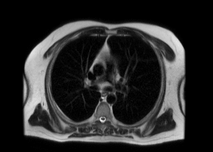
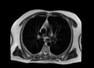
Lungs and pleura: No pleural effusions. Limited evaluation of the lungs demonstrates no abnormal signal characteristics.
Visualized upper abdominal organs: Unremarkable.
IMPRESSION: Limited exam, as noted above. Dilated cardiomyopathy, with moderately enlarged left ventricular chamber size and severely depressed left ventricular systolic function. Ejection fraction equals 16%. There is diffuse hypokinesia of the left ventricular walls, with akinesia of the apex and septum. Mild nonspecific late gadolinium enhancement involves the mid septum as well. Normal chamber size and systolic motion of the right ventricle. Normal size left and right atria. No significant cardiac valvular disease. Normal caliber aorta and main pulmonary artery. No evidence of pericardial or pleural effusions.