
MR HEAD AND ORBITS WITH AND WITHOUT CONTRAST
HISTORY: Headaches – Blurry Vision
COMPARISON: 9/12/2013
TECHNIQUE: Multiplanar images of the head were obtained at 1.5 Tesla prior to and following administration of 6 mL of Gradavist intravenous contrast. Thin sections through orbits were also obtained.
FINDINGS:
HEAD:: There are mild hypertrophic degenerative changes of the acromioclavicular joint. There is mild narrowing of the subacromial space. There is minimal fluid within the subacromial subdeltoid bursa.
Brain: There is no infarct, hemorrhage, mass or hydrocephalus. There is no enhancing lesion.
Diffusion Images: No evidence of acute ischemic injury
Pituitary: There is no sellar lesion
Vasculature: Normal vascular flow voids are demonstrated
LACS: The internal auditory canals are normal
Sinuses: There is a diffuse patter of mucosal thickening involving the paranasal sinuses. The mastoid air cells are clear.
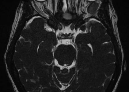
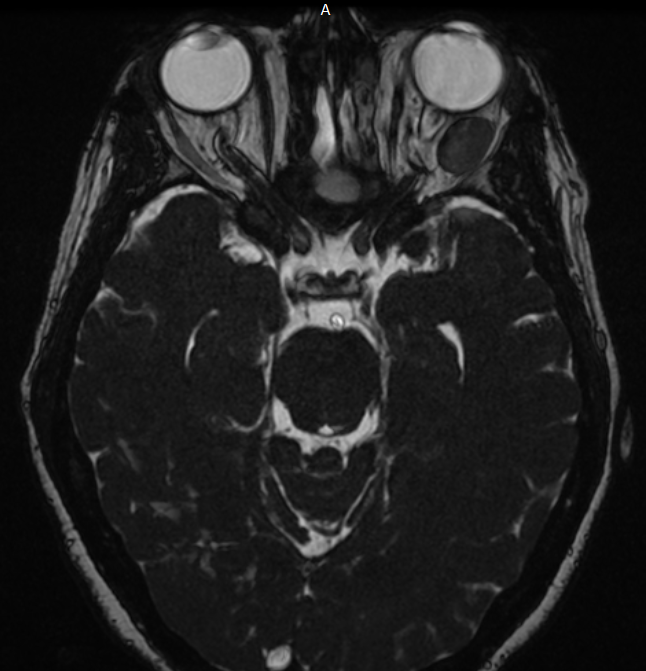
ORBITS: As noted on prior studies, there is an oval smoothly marginated intraconal mass of the left orbit, just posterior to the globe and superior and latera to the left optic nerve. Compared to the prior study, the lesion has increased slightly in size, now measuring 17 mm in greatest length, compared to 14 mm previously. The lesion enhances uniformly with contrast, suggestive of a hemangioma. The globes are symmetric and normal in appearance. There is some distortion of the left lateral rectus muscle by the left orbital mass, but the extraocular muscles otherwise symmetric and normal in appearance. The optic nerves and optic chiasm are unremarkable.
IMPRESSION: Slowly progressive left orbital mass, now measuring 17 mm in greatest length, just posterior to the left globe and lateral to the left optic nerve, nonspecific in appearance but suggestive of an orbital hemangioma