
 MR HEAD
HISTORY: Evaluate for intra- and extracranial hemorrhage
COMPARISON: Ultrasound 06/20/2023
TECHNIQUE: MR Brain Multiplanar images of the head were obtained at 1.5 Tesla without IV contrast.
FINDINGS:
Brain: There is no infarct, hemorrhage, mass or hydrocephalus.
Diffusion Images: No evidence of acute ischemic injury.
Pituitary: There is no sellar lesion.
IACS: The internal auditory canals are unremarkable.
Vasculature: Normal vascular flow voids are demonstrated.
Sinuses: The paranasal sinuses appear unremarkable.
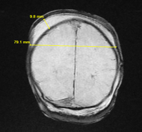
Bones and Soft Tissues: There is a parietal scalp hematoma crossing the midline larger on the right than left. This measures approximately 8 cm in diameter and up to 11 mm in thickness. This extends into the left frontotemporal region. There appears to be an overlying left parietal scalp hematoma. There is no definite calvarial fracture.
IMPRESSION: Right larger than left scalp hematoma crossing the midline. This could represent a subgaleal hematoma or subcutaneous scalp hematoma.
No definite subdural or epidural hematoma.