


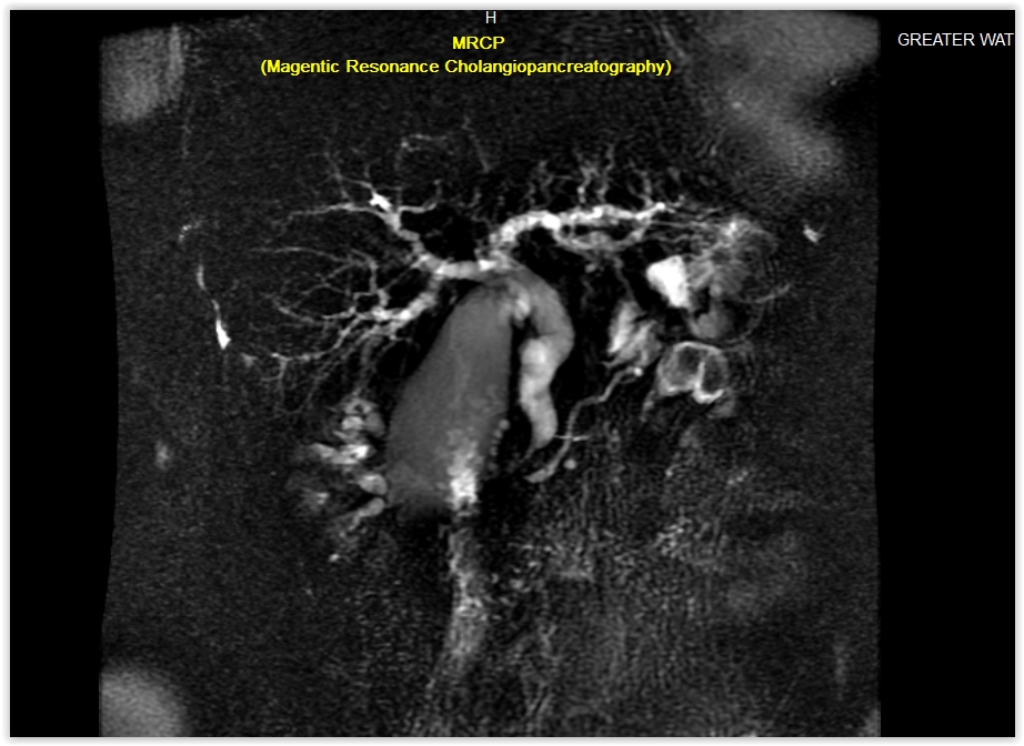
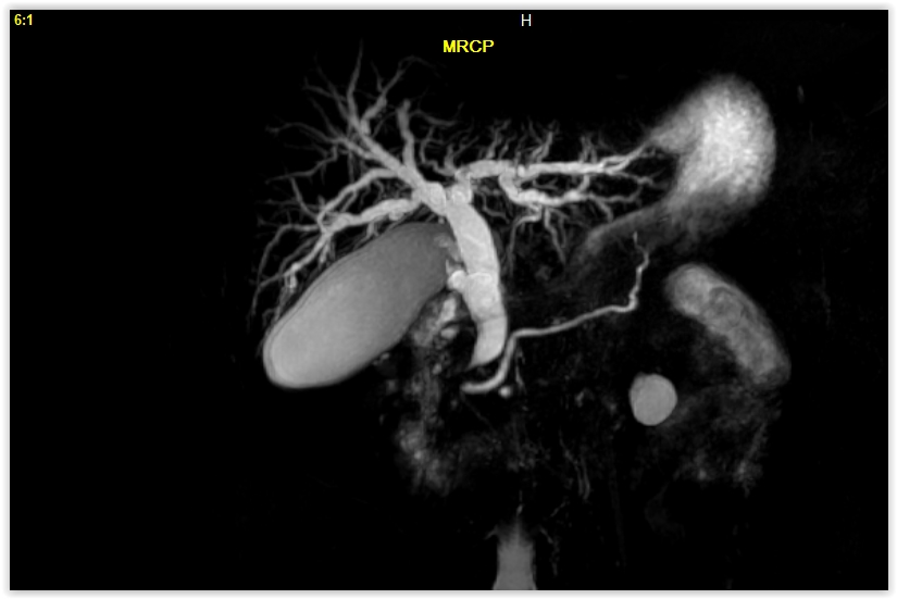
Magnetic Resonance Cholangiopancreatography – MRCP Case Study
History: Female patient had history of abdominal pain from bile duct obstruction
Comparison Study: CT of the abdomen which was done in November 2015 and abdominal ultrasound in 2007
MRCP Exam Technique: Multiplanar images of the biliary tree were obtained without intravenous contrast using the GWIC 1.5T MRI system
Findings:
- The gallbladder is moderately distended; no gallstones, wall thickening; or pericholecystic fluid is seen.
- The common bile duct is also moderately distended, measuring up to approximately 11 mm in diameter. The distal common bile duct tapers in caliber in the pancreatic head, to approximately 1 mm in diameter near the ampulla. No CBD stone is seen.
- There is moderate diffuse intrahepatic biliary dilatation seen in the liver.
- The pancreas appears normal. No pancreatic mass is seen. The pancreatic duct is not dilated.
- The spleen appears normal.
- The adrenal glands appear normal.
- There is no renal mass or hydronephrosis seen in the kidneys. There are two left renal cortical cysts measuring 2.3 cm and 0.6 cm.
- No evidence of aortic aneurysm is seen.
- No abdominal lymphadenopathy is present.
Impression: Smooth narrowing of the distal common bile duct. There is intrahepatic and extrahepatic biliary dilatation. No distal CBD stone or pancreatic head mass is seen. Findings are suggestive of a biliary stricture. The biliary tree is similar compared to the CT of 11/2015 although was not dilated on the examination from 2007. There is no evidence of acute pancreatitis. The pancreatic duct is not dilated. No gallstones are seen.