

MR ABDOMEN KIDNEY W WO
(Reason for Exam: MR Abdomen Kidney w wo) RENAL MASS
HISTORY: RENAL MASS
COMPARISON: None
TECHNIQUE: Multiplanar images of the abdomen were obtained at 1.5 Tesla prior to and following administration of 15 mL of Dotarem intravenous contrast. During this public health emergency, we are using enhanced sterilization processes, social distancing measures and PPE for your protection.
FINDINGS:
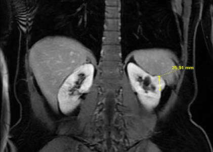
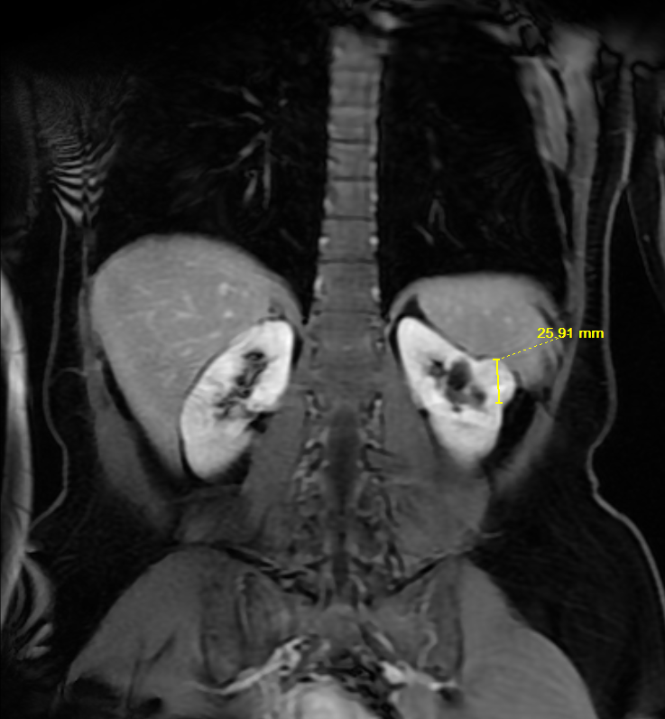
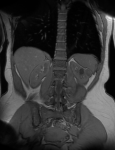
Kidneys: There is a complex exophytic cortical lesion projecting laterally from the mid-zone of the left kidney measuring 2.9×2.2×2.6 cm. This lesion is heterogeneously enhancing and suspicious for a renal malignancy. The mass does not involve the renal hilum. No additional enhancing renal lesion is seen. There are benign bilateral renal cortical cysts. The largest left renal cyst is seen within the mid to upper pole, measuring 1.9 cm in length. There is normal symmetric cortical enhancement of both kidneys. The renal veins are patent. No evidence in the perinephric soft tissues.
Adrenal glands: the adrenal glands appear normal.
Liver: deliver appears normal.
Gallbladder: The patient is status post cholecystectomy.
CBD: the common bile duct is normal in caliber.
Pancreas: the pancreas appears normal.
Spleen: the spleen appears normal.
Aorta: no evidence of aortic aneurysm or dissection is seen.
Lymphatics : no abdominal lymphadenopathy is present.
IMPRESSION:
Enhancing left renal cortical mass suspicious for renal cell carcinoma. This may also represent oncocytoma. No evidence of metastatic disease. No adenopathy.