
 HISTORY: CIRRHOSIS OF LIVER
COMPARISON: CT Abdomen 7/29/20
TECHNIQUE: Multiplanar images of the abdomen were obtained at 1.5 Tesla prior to and following administration of IV contrast. During this public health emergency, we are using an enhanced sterilization processes, social distancing measures and PPE for your protection.
CONTRAST: 9 mL of Eovist intravenous contrast.
FINDINGS:
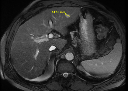
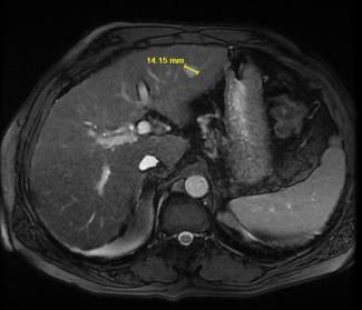
Liver: the liver is heterogeneous and nodular in contour consistent with cirrhosis. There is a T2 hyperintense lesion in the lateral segment measuring 1.4 cm in length. This demonstrates peripheral globular enhancement with delayed filling in consistent with a benign hemangioma. There is a 1.1 cm T2 hyperintense lesion along the liver dome within the anterior segment which is nonenhancing and is consistent with a benign cyst. There is an additional nonenhancing lesion measuring 8 mm inferiorly within the posterior segment also consistent with a benign cyst. No suspicious enhancing liver lesion is seen. The portal vein is patent.
Gallbladder: the gallbladder appears normal, without gallstones, wall thickening or pericholecystic fluid.
CBD: the common bile duct is normal in caliber.
Pancreas: the pancreas appears normal.
Spleen: The spleen appears normal.
Adrenals: the adrenal glands are normal.
Kidneys: there is no renal mass or hydronephrosis.
Aorta: no evidence of aortic aneurysm or dissection is seen.
Lymphatics: no abdominal lymphadenopathy is present.
IMPRESSION:
Cirrhosis. No evidence of hepatic malignancy. No gallstones or biliary obstruction.
