
HISTORY: LEFT HEPATIC HEMANGIOMA
COMPARISON: July 25, 2016 and February 26, 2010
TECHNIQUE: Multiplanar images of the abdomen were obtained at 1.5 Tesla prior to and following administration of IV contrast.
CONTRAST: 20 mL of Dotarem intravenous contrast
FINDINGS:
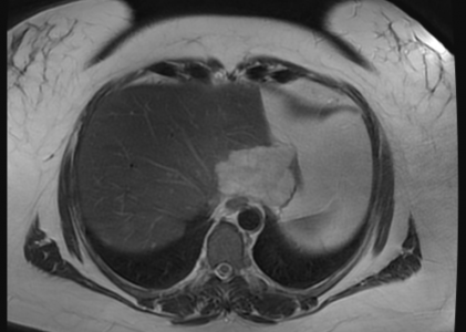
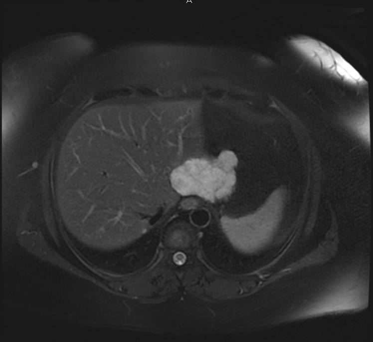
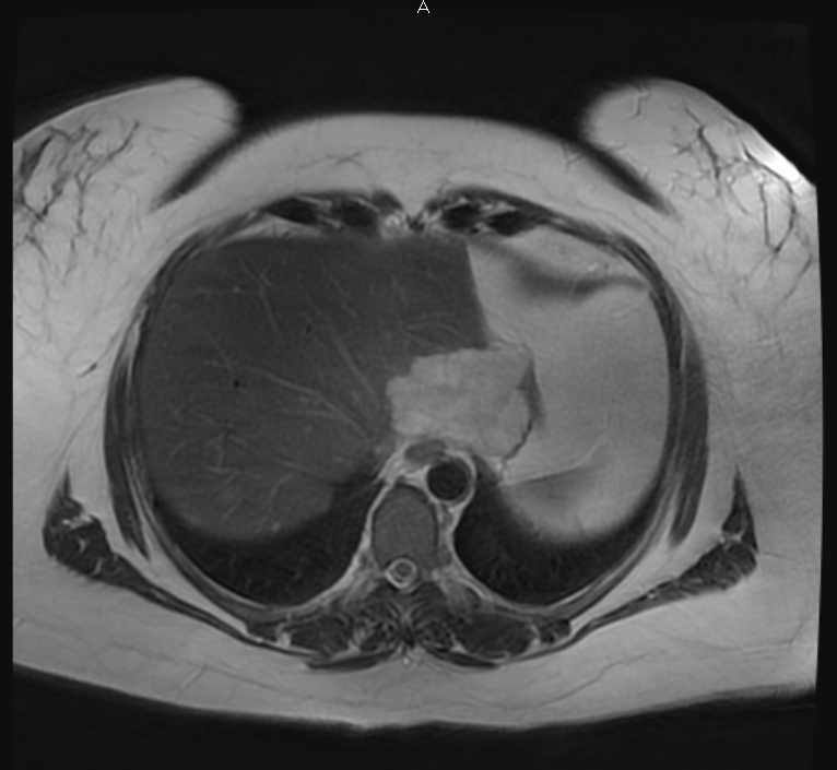
Liver: Again seen are several lesions in the liver. They show the typical bright appearance of hemangiomas on T2-weighted imaging. The larger lesions also show he typical gradual globular contrast enhancement of hemangiomas. The largest lesion posteriorly in the left lobe of the liver measures 6.6 cm, not significantly changed from the size in 2010. The second largest lesion posteriorly in the right lobe of the liver measured 7.0 cm in 2010, but now measures only 3.3 cm. Several other smaller hemangiomas are not significantly changed.
Gallbladder: The gallbladder appears normal, without gall stones, wall thickening or pericholecystic fluid.
CBD: The common bile duct is normal in caliber.
Pancreas: The pancreas appears normal.
Spleen: The spleen appears normal.
Adrenal Glands: The adrenal glands appear normal.
Kidneys: The kidneys appear normal. No mass or hydronephrosis is seen.
Aorta: No evidence of aortic aneurysm or dissection is seen.
Lymphatics: No abdominal lymphadenopathy is present.
IMPRESSION:
Multiple benign liver hemangiomas. Most of them are unchanged, although the second largest lesion, in the right lobe of the liver, has partially involuted since 2010.