


MR Bilateral Breasts With and Without IV Contrast Case Study
History: Newly diagnosed breast cancer
Technique: Multiplanar images of the breasts were obtained at 1.5 Tesla on a dedicated breast coil prior to and following administration of 15 mL of Omniscan intravenous contrast.
Comparison: Mammogram 3/27/2015
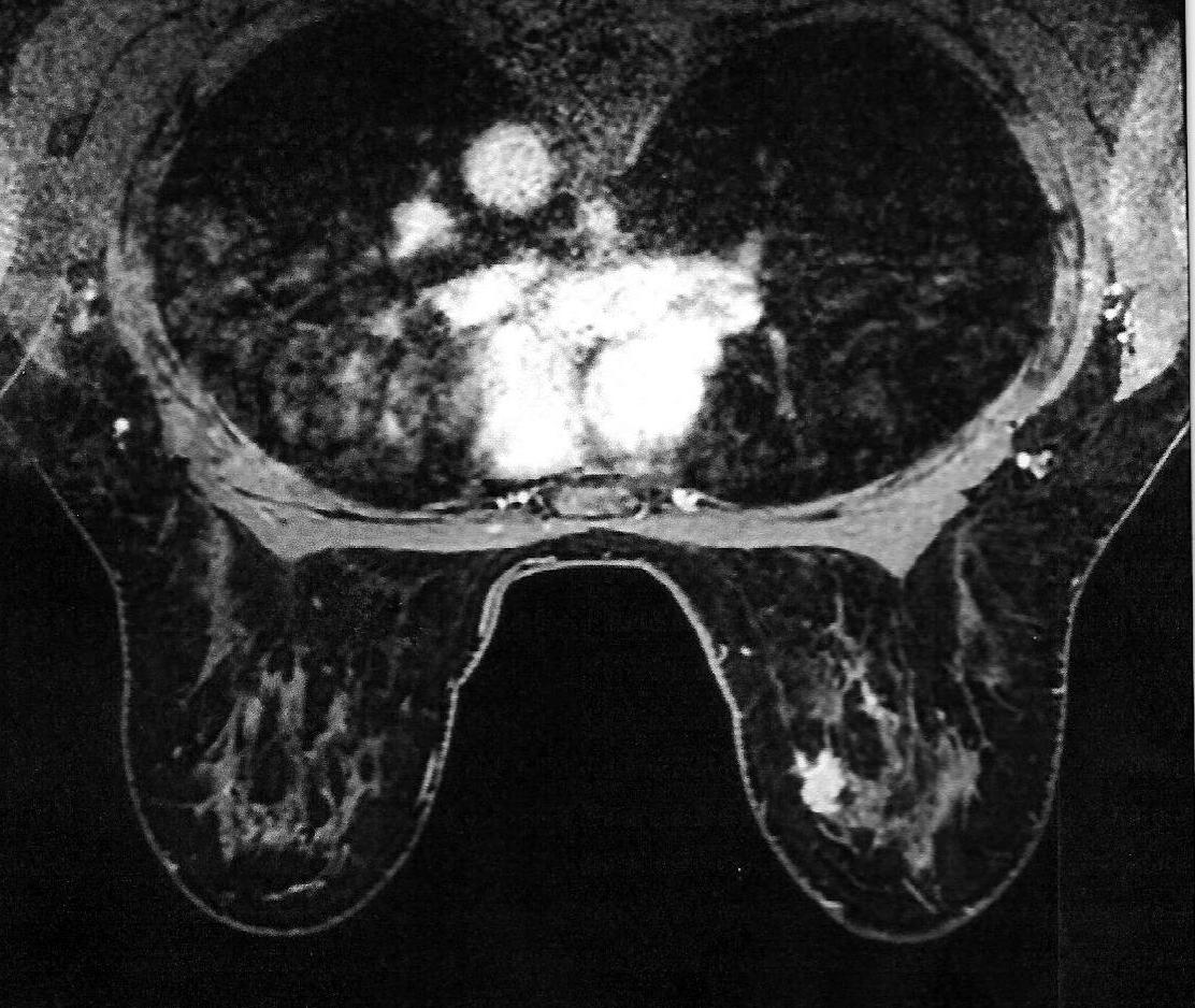
Findings: There is a stereotactic biopsy marker at the 2:00 position of the right breast, approximately 5.5 cm deep to the nipple. Immediately surrounding the clip is an area of heterogeneous parenchymal enhancement, suspicious for malignancy measuring 2.4 x 1.3 cm in transverse diameter. The borders are irregular. The lesion is hypervascular on early phase postcontrast images. There is no axillary adenopathy. No additional breast lesion is seen bilaterally.
This study was evaluated using a computer aided detection software program.
Impression: Enhancing lesion around the stereotactic biopsy clip suspicious for malignancy. No evidence of metastatic disease is otherwise seen.
ACR BIRADS: #5: Highly suggestive of malignancy.