

 HISTORY: VASCULAR DEMENTIA
COMPARISON: 3/16/2020
TECHNIQUE: Multiplanar images of the head were obtained at 1.5 Tesla without IV contrast.
FINDINGS:
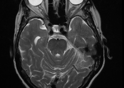
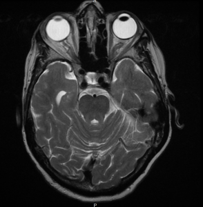
Brain: There is a small old right occipital lobe infarct. There is mild diffuse cerebral atrophy. Scattered chronic small vessel ischemic changes are present throughout the periventricular and subcortical white matter bilaterally. No intracranial mass or evidence of recent hemorrhage. There are scattered ares of hemosiderin deposition, related to old sites of hemorrhage associated with old ischemic areas.
Diffusion Images: No evidence of acute ischemic injury.
Pituitary: There is no sellar lesion.
IACS: The internal auditory canals are unremarkable.
Vasculature: Normal vascular flow voids are demonstrated.
Sinuses: The paranasal sinuses appear unremarkable.
IMPRESSION: Old right occipital lobe infarct. Atrophy and chronic small vessel ischemic disease, similar to prior exam. No acute infarct or other acute intracranial process.