
HISTORY: MULTIPLE SCLEROSIS 
TECHNIQUE: multiplanar images of the head were obtained at 1.5 Tesla prior to and following administration of 17 mL of Dotarem intravenous contrast. During this public health emergency, we are using enhanced sterilization processes, social distancing measures and PPE for your protection.
COMPARISON: MRI brain 11/17/2020
FINDINGS:
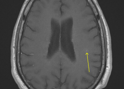
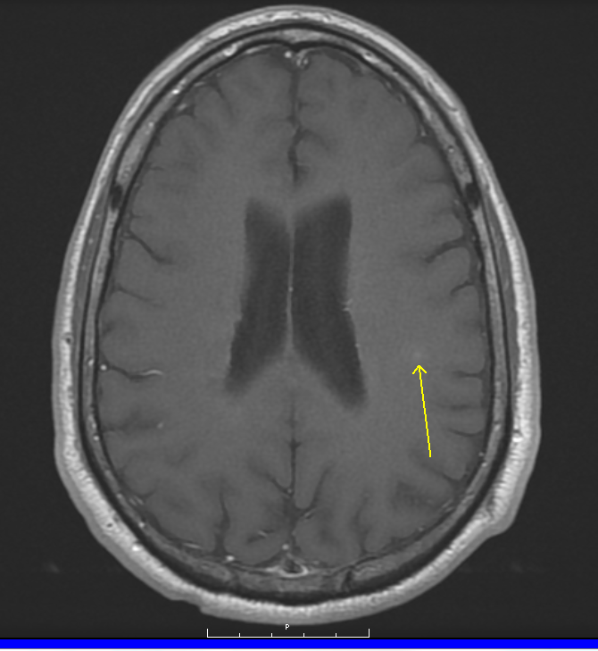
Brain: Redemonstrated are numerous foci of periventricular and subcortical FLAIR signal alteration several oriented perpendicular to the long axis. There is a FLAIR signal alteration in the left middle cerebellar preduncle is unchanged. There is a new focus of FLAIR signal alteration in the left inferior frontal gyrus demonstrating very subtle enhancement following the administration of contrast. No additional enhancing lesions are identified. No intracranial mass, mass effect or midline shift. No acute territorial infarct or intracranial hemorrhage. The ventricles are unchanged in size and configuration.
Pituitary: There is no sellar lesion.
IACS: The internal auditory canals are unremarkable.
Vasculature: Major intracranial flow voids at the skull base are patent.
Sinuses: Minor paranasal sinus mucosal thickening. Mastoid air cells are clear.
IMPRESSION:
New white matter lesion left frontal lobe demonstrating with subtle enhancement following the administration of contrast suspicious for active demyelination. Otherwise, white matter lesions appear unchanged.