
HISTORY: Concern for new ischemic stroke
COMPARISON: CT head 5-6-21, 5-7-21 and 5-15-21
TECHNIQUE: Multiplanar images of the head were obtained at 1.5 Tesla prior to and following administration of 15 mL Dotarem intravenous contrast. During this public health emergency, we are using enhanced sterilization processes, social distancing measures and PPE for your protection.
FINDINGS:
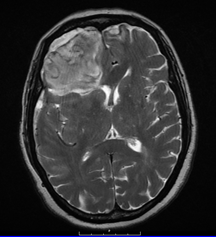
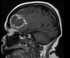
Brain: Redemonstrated is a right frontal lobe intraparenchymal hematoma similar in size compared to the most recent CT allowing for differences in modality. On the current study, the hematoma measures approximately 6.4 x 4.8 cm. There is no significant enhancement. No evidence of intraventricular extension. There is a slight holohemishperic subdural hematoma measuring 7 mm in greatest transverse thickness along the right frontal convexity and extending along the falx. Mass effect contributes to similar appearing effacement of the right frontal lobe sulci and frontal horn of the right lateral ventricle. Unchanged 6 mm is leftward midline shift.
There is a punctate focus of diffusion restriction in the right cerebellar hemisphere.
Areas of periventricular and subcortical FLAIR signal alteration are compatible with sequelae of chronic small vessel ischemic change. Generalized atrophy is associated with concomitant enlargement of the sulci and ventricles.
Pituitary: There is no sellar lesion.
IACS: The internal auditory canals are unremarkable.
Vasculature: Major intracranial flow voids at the skull base are patent.
Sinuses: There is a minor paranasal sinus mucosal thickening with a small mucous retention cyst versus polyp in the left maxillary sinus. Paranasal sinuses and mastoid air cells are otherwise clear.
IMPRESSION:
Right frontal lobe intraparenchymal hematoma and holohemispheric right-sided subdural hematoma mass effect and leftward midline shift appears similar compared to the most recent CT. No definite underlying mass. Short interval follow-up recommended to ensure resolution.
Punctate acute subacute infarct in the right cerebellar hemisphere.