
HISTORY: Stroke evaluation
COMPARISON: 1/23/20
TECHNIQUE: Multiplanar images of the head were obtained with 1.5 Tesla without IV contrast.
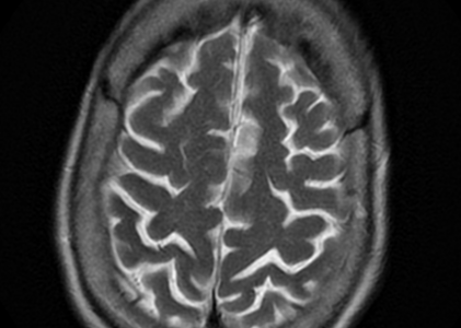
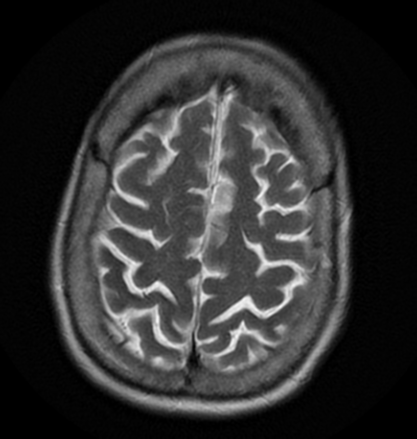
FINDINGS:
Brain: There is no infarct, acute hemorrhage, mass or hydrocephalus. There are mild atrophic and small vessel ischemic changes. There is mild cerebellar atrophy. There are old bilateral thalamic lacunar infarcts. There are small foci of old hemorrhage in the deep basal ganglia/thalami, bilaterally, as well as in the left cerebellar hemisphere.
Diffusion Images: No evidence of acute ischemic injury.
Pituitary: There is no sellar lesion.
IACS: The internal auditory canals are unremarkable.
Vasculature: Normal vascular flow voids are demonstrated.
Sinuses: The paranasal sinuses appear unremarkable.
IMPRESSION: No acute infarct. Atrophic and small vessel ischemic changes. Old lacunar infarcts. Tiny, scattered areas of hemorrhage.
