MR liver iron quantification is a non-invasive means of measuring liver iron concentration, a key indicator in the management of patients with hemochromatosis (primary or secondary). Hemochromatosis is characterized by a progressive increase in total body iron stores, with abnormal iron deposition in multiple organs. Primary hemochromatosis is a genetic disorder, whereas secondary hemochromatosis can be the result of a variety of disorders, most commonly chronic hemolytic anemia.
An advantage apart from being non-invasive is that sampling occurs in a large cross-section of the liver as opposed to a biopsy, which typically represents 1/50,000 of the whole liver.
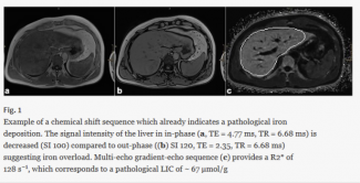
There is an easy way to get an idea of first impression of a possible iron overload in the liver with MRI: nearly every MRI protocol of the liver integrates a chemical shift sequence, i.e., in-and opposed-phase. Iron leads to decreased signal intensity on in-phase images compared with the opposed-phase which in turn should alert on a possible iron overload disease. An example is provided in Fig.1.