
HISTORY: Rectal cancer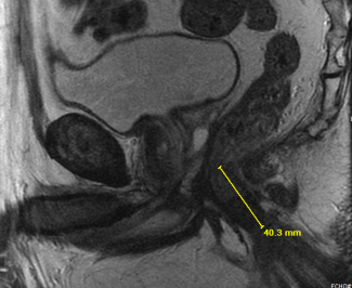
COMPARISON: None
TECHNIQUE: Multiplanar images of the pelvis were obtained at 1.5 Tesla without IV contrast. Sequences: Large FOV axial T1 and sagittal T2-weighted images. Small FOV axial, oblique axial, coronal, oblique coronal and sagittal T2-weighted images. Axial diffusion-weighted images. Axial contrast-enhanced T1-weighted images. The image quality is adequate.
FINDINGS:
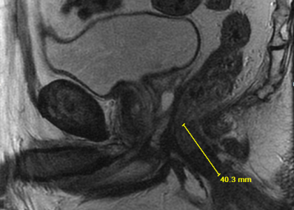
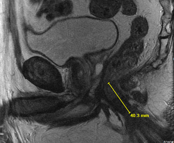
TUMOR LOCATION AND CHARACTERISTICS: Tumor location from anal verge: Low 0-5.0 cm Anal verge to distal tumor margin: 4 cm Tumor at or below the puborectalis sling: No Distance of lowest extent of tumor from top of anal sphincter: 0 cm Relationship to the anterior peritoneal reflection: Below Craniocaudal length of the tumor:4.7 cm Clock face of tumor: 2 o’clock to 10 o’clock Polypoid/annular/semiannular: Semiannular Mucinous: No
EXTRAMURAL DEPTH OF INVASION AND MR T-CATEGORY Extramural depth of invasion (Use 0 mm for TI or T2 tumor): <5 mm T-Category: T3 For low rectal tumors (maximum tumor depth at or below the puborectalis sling): 0
RELATIONSHIP OF THE TUMOR TO MESORECTAL FASCIA (MRF) Shortest distance of the definitive tumor border to the MRF is: 0 mm at 6 o’clock Are there any tumor spiculations closer to the MRF? No
EXTRAMURAL VENOUS INVASION Extramural venous invasion (EMVI): Absent
MESORECTAL LYMPH NODES AND TUMOR DEPOSITS
Any suspicious mesorectal lymph nodes: Yes. The most suspicious node/tumor deposit is at the tumor with minimum distance 5 mm from the MRF at 4 o’clock.
EXTRAMESORECTAL LYMPH NODES. Any suspicious extramesorectal lymph nodes: No
Is the IMA node station in the field of view: Yes. The nodes are not suspicious.
OTHER FINDINGS (COMPLICATIONS, METASTASES, LIMITATIONS)
Urinary Bladder. The urinary bladder appears normal, without stones or wall thickening. Prostate: The prostate is normal in size and signal.
Seminal Vesicles: The seminal vesicals are symmetric bilaterally and appear unremarkable. Abdominal Wall: No abdominal wall or inguinal hernia is seen.
Bones: The bones appear unremarkable.
IMPRESSION:
MRI rectal cancer T-Category is: 13 Maximum EMD of invasion is: <5 mm Minimum tumor to MRF distance is: 0 mm Low rectal tumor component: Yes Mesorectal nodes/tumor deposits: Suspicious
EMVI: Absent Extramesorectal Nodes: Negative