MR PELVIS WITH AND WITHOUT IV CONTRAST
HISTORY: PERSISTENT RECTAL PAIN – RESTAGE RECTAL CANCER
COMPARISON: 3/14/2018
TECHNIQUE: Multiplanar images of the right pelvis were obtained at 1.5 Tesla prior to and following administration of 16 mL of Dotarem intravenous contrast.
FINDINGS:
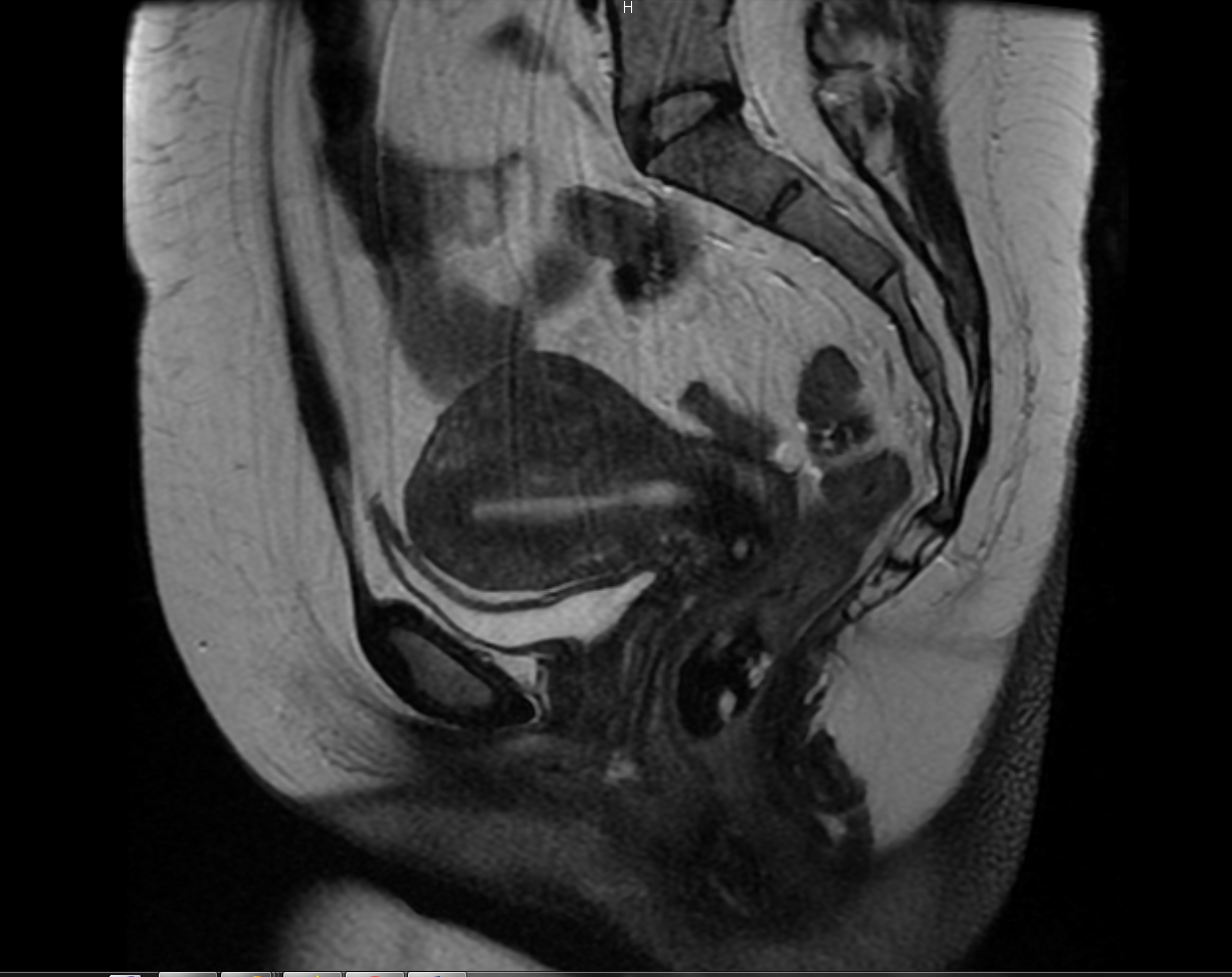
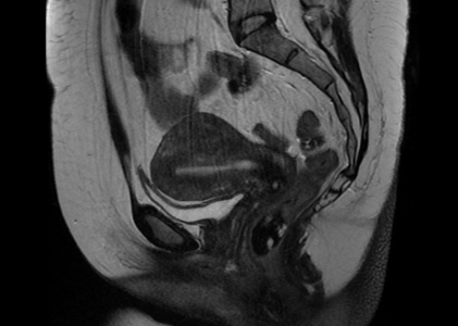
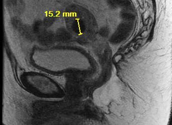
GI tract: Since the last exam, the patient has undergone AP resection. There is a left lower quadrant colostomy. No residual or recurrent pelvic mass. There is some prolapse of the pelvic floor soft tissue structures, with fluid collecting inferior to the cervix posteriorly.
Uterus: The uterus is normal in size, but contains multiple small fibroids, largest in the anterior uterine body measuring 1.5 cm in diameter. There are several small cervical nabothian cysts.
Endometrium: The endometrium is normal in thickness.
Ovaries: The ovaries appear normal. No masses or cysts are seen.
Urinary Bladder: The urinary bladder appears normal, without stones or wall thickening.
Abdominal Wall: No abdominal wall or inguinal hernia is seen.
Lymphatics: No pelvic lymphadenopathy is present.
Bones: The bones appear unremarkable.
IMPRESSION: Status post AP resection, with a left lower quadrant colostomy. No residual or recurrent pelvic mass or adenopathy. There is inferior prolapse of the pelvic floor, with some fluid collecting inferiorly, but no associated mass.