

 HISTORY: RIGHT KNEE PAIN
COMPARISON: None
TECHNIQUE: Multiplanar images of the right knee were obtained at 1.5 Tesla without IV contrast.
FINDINGS:
LIGAMENTS:
ACL: There is mucinous degeneration of the ACL proximally, without a discrete ligament tear.
PCL: The posterior cruciate ligament is intact.
MCL: The medial collateral ligament is intact.
LCL: The lateral collateral ligament is normal.
MENISCI:
Medical Meniscus: The medial meniscus is intact.
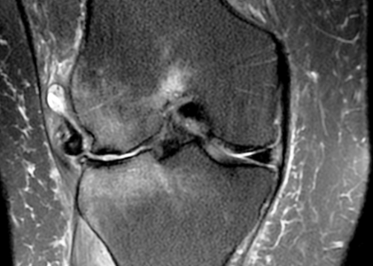
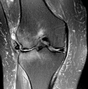
Lateral Meniscus: There are chronic tears of the anterior horn and body of the lateral meniscus, with displacement of the body of the meniscus laterally from the joint space.
BONES: There is severe osteoarthritis of the lateral femorotibial joint, with extensive loss of articular cartilage along the lateral femoral condyle and lateral tibial plateau. There is some remodeling of the bony articular surfaces, with subchondral sclerosis on both sides of the joint and subchondral bone marrow edema within the lateral femoral condyle and lateral tibial plateau. No evidence of acute fracture. There are prominent lateral marginal osteophytes, with smaller osteophytes medially and small spurs encroaching on the intercondylar notch. There are degenerative changes of the patellofemoral joint, with erosions of articular cartilage primarily involving the lateral patellar facet and slight lateral subluxation of the patella.
SOFT TISSUES: The quadriceps and patellar tendons are intact. There is a moderate knee joint effusion, with a 5 cm medial popliteal cyst.
IMPRESSION: Chronic degenerative tears of the body and anterior horn of the lateral meniscus, with lateral displacement of the meniscus from the joint space and severe osteoarthritis of the lateral femorotibial joint.