
HISTORY: acute CVA
COMPARISON: None.
TECHNIQUE: Two-dimensional time-of-flight images and contrast enhanced MRA images of the neck were obtained at 1.5 Tesla prior to and following administration of 16 mL of Dotarem intravenous contrast. During this public health emergency, we are using enhanced sterilization processes, social distancing measures and PPE for your protection.
FINDINGS:
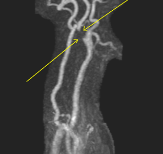
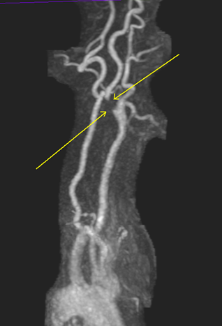
Carotid Arteries: There is signal dropout over an 8 mm segment involving the proximal aspect of the left internal carotid artery with slow noted within the remainder of the internal carotid artery into the neck. This is suggestive of a severe stenosis of 90-99%. The right internal carotid artery in the neck is patent. There is a moderate focal stenosis of the cavernous and precavernous portion of the right internal carotid artery. The common carotid arteries are patent.
Vertebral Arteries: No occlusion or significant stenosis is seen. There is normal direction of blood flow.
IMPRESSION:
Findings most significant for a severe right proximal ICA stenosis in the 90-99% range.
There is likely a small to moderate focal stenosis of the cavernous and precavernous portion of the right internal carotid artery.
Direct measurements of the distal internal carotid diameters were utilized as the denominator for stenosis measurement.
PQRS 195: CPT II: 3100F: Carotid imaging study report includes direct or indirect reference to measurements of the distal internal carotid diameter as the denominator for stenosis measurement.