


MRI Abdominal Hepatitis C Case Study
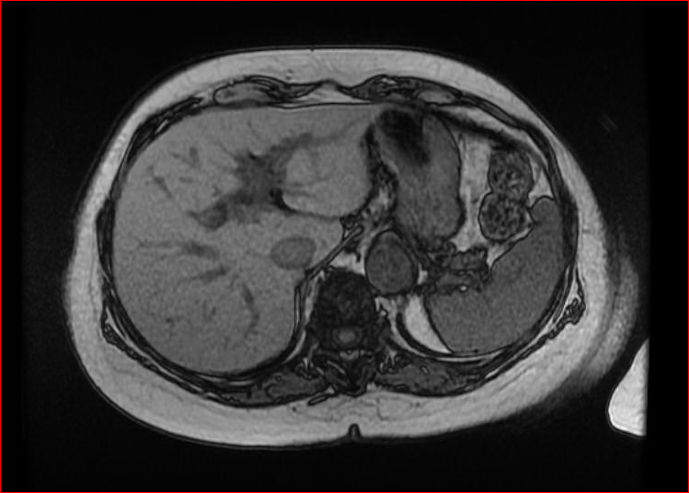
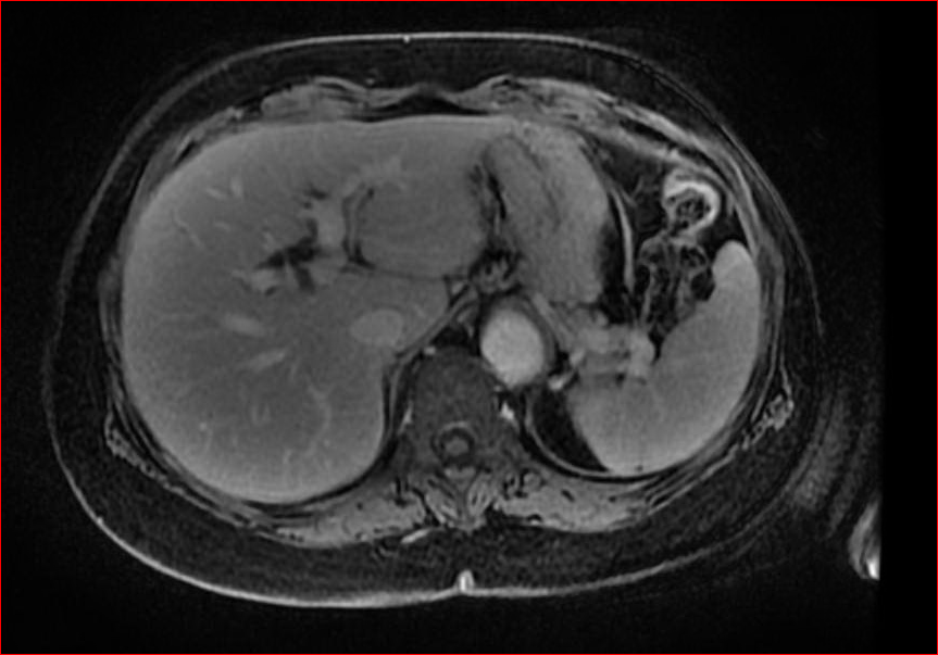
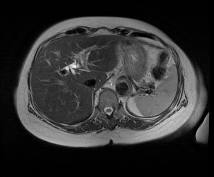
MRI screening for Hepatitis C provides the most sensitivity for soft tissue, allowing for easier detection of liver tumors. Below are 4 of the different views that are obtained for the study of the liver. The patient came for the MRI exam for history of anemia and hepatitis C. The MRI Abdominal Hepatitis C Case Study procedure included multi-planar images of the abdomen obtained with and without IV contrast on our 1.5 Tesla MRI machine. The comparison Ultrasound scan was done on 4/15/2016.
MRI Exam Findings:
History: Anemia, Hepatitis C
Comparison: Ultrasound 4/15/2016
Technique: Multiplanar images of the abdomen were obtained at 1.5 Tesla prior to and following administration of IV contrast.
Contrast: Eight mL of Gadavist intravenous contrast.
FINDINGS:
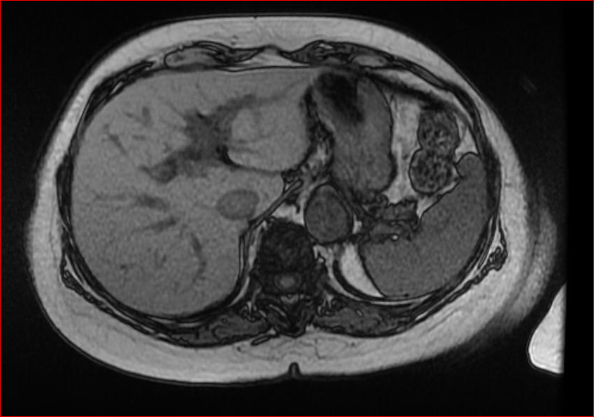 Liver: There is a heterogeneous texture of the liver, with a small nonspecific linear focus of hypervascularity just above the gallbladder fossa at the junction of the left and right lobes. No discrete hepatic mass. There is a minor pattern of intrahepatic biliary ductal dilation, without evidence of an obstructing lesion. The calcified granulomata demonstrated in the liver on the ultrasound exam are not visible on the MR study.
Gallbladder: The patient is status post cholecystectomy.
 CBD: The common bile duct measures 9 mm in caliber, most likely representing physiologic dilatation following cholecystectomy.
Pancreas: The pancreas appears normal.
 Spleen: The spleen appears normal.
Adrenal Gland: The adrenal glands appear normal.
Kidneys: The kidneys appear normal. No mass or hydronephrosis is seen.
Aorta: No evidence of aortic aneurysm or dissection is seen.
Lymphatics: No abdominal lymphadenopathy is present.
IMPRESSION:
 Heterogeneous liver parenchyma consistent with a history of hepatitis C, but without evidence of a discrete mass. Mild dilation of the intrahepatic and extrahepatic ducts is probably physiologic, related to prior cholecystectomy. Calcified hepatic granulomata demonstrated on recent ultrasound exam are not visible on the MR exam.