


MRI Ankle Case Study 2
 Our case study of the month is an MRI scan of the left ankle. The patient presented with left ankle pain following an injury. The MRI Ankle Case Study procedure included multi-planar images of the left ankle obtained without IV contrast on our 1.5 Tesla MRI machine. There was no comparison scan.
MRI Exam Findings:
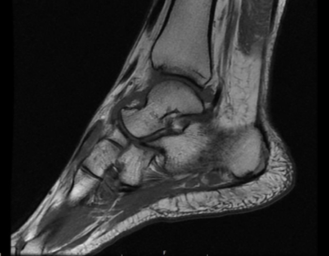 Bones: There is a small area of bone marrow edema within the distal cuboid bone. No discrete cortical fracture is seen; findings most consistent with a bone bruise. No additional areas of abnormal bone marrow signal are seen. There are mild degenerative changes of the talo-navicular joint.
Tendons: The flexor and extensor tendons are intact. The Achilles tendon and plantar fascia are intact.
Ligaments: There is abnormal signal within the tibiocalcaneal ligament, consistent with a mild ligamentous sprain. There is no evidence of ligamentous rupture. The distal tibiofibular ligaments, the tibiocalcaneal ligament and the lateral collateral ligament complex are intact.
Soft Tissues: There is mild subcutaneous edema medially and laterally. No mass or abnormal fluid collection is seen. No significant joint effusion is present.
Impression: Mild sprain of the tibiocalcaneal ligament. No ligamentous rupture.
For more information on MRI imaging services at Greater Waterbury Imaging Center, visit the blog post on Sprains and Strains and our clinical section of the website.