MRI Cervical Spine Case Study
Our case study of the month is an MRI scan of the cervical spine. The patient presented with neck pain, arm numbness and had been in a car accident 1 week prior to the imaging exam. The MRI Cervical Spine Case Study procedure included axial and sagittal images of the cervical spine which were obtained on our 1.5 Tesla MRI machine.
MRI Exam Findings
Indications: Neck Pain – Radiculopathy
Technique: Axial and sagittal images of the cervical spine were obtained at 1.5 Tesla
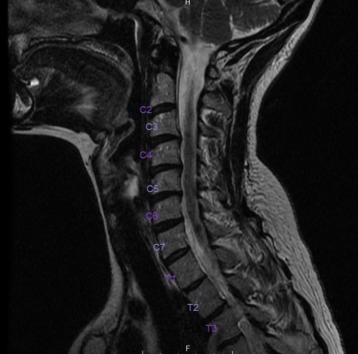
Findings: There are mild degenerative disk changes from C3 through C7. At C3-4 and C4-5, there are very small broad central protrusions causing mild stenosis. At C5-6, there is a bulge causing minor stenosis. There is mild right-sided neural foraminal narrowing at C4-5 secondary to uncinate hypertrophy. At C6-7, there is a minor bulge. At T2-3, there is a minor central protrusion. No intradural abnormalities are noted. No discrete vertebral body lesions are seen. The remaining neuroforamina are patent.
Impression: There is mild spondylosis and small bulges and protrusions superimposed on a congenitally narrow canal, causing mild stenosis, most significant at C3-4 and C4-5, as discussed above.