


MRI Chest Case Study With and Without Contrast
History
Elastofibroma dorsi is a benign soft tissue pseudotumor which is uncommon and frequently located at the lower pole of the scapula, deep to the serratus anterior, and it is often attached to the periosteum of the ribs. Patients with elastofibroma dorsi present with a long history of swelling and, occasionally, pain and discomfort. As it is often bilateral, the contralateral site should be evaluated. See this reference for more detail – https://en.wikipedia.org/wiki/Elastofibroma_dorsi

Indications: Chest wall mass
Comparison: None
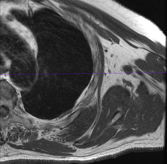
Technique: Coronal, axial and oblique sagittal views of the left upper chest were obtained at 1.5 Tesla, without and with contrast administration. 10 cc Gadavist injected intravenously.
Findings: Superficial to the posterior lareral wall of the left upper chest, deep to the scapula and deep to the left serratus anterior and latissimus dorsi muscles, there is a striated oval non-encapsulated 7 cm soft tissue mass, with MR characteristics consistent with an elastofibroma dorsi. There is only faint enhancement of this lesion after contrast administration. The ribs of the left posterior chest wall are unremarkable. The scapula is normal in appearance. The muscles of the rotator cuff are unremarkable.
Impression: 7 cm mass of the left chest wall posterolaterally, deep to the scapula has imaging characteristics consistent with a typical elastofibroma dorsi.