

 HISTORY: Atypical facial pain
COMPARISON: None
TECHNIQUE: Multiplanar images of the orbits were obtained at 1.5 Tesla prior to and following administration of 10 ml of Dotarem intravenous contrast. During this public health emergency, we are using enhanced sterilization processes, social distancing measures and PPE for your protection.
FINDINGS:
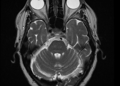
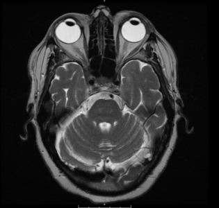
Orbits: At the level of the right cavernous sinus and right orbital apex, there is an enhancing soft tissue mass extending through the right superior orbital recess and right optic canal along the right orbital roof posteriorly, superior to the right superior rectus muscle. This finding could represent a meningioma, but has a relatively aggressive appearance and could represent metastatic tumor. The lesion extends over an AP length of 3.5 cm, from the cavernous sinus into the right orbit along the orbital roof, to the level of the posterior margin of the globe and measures 8 mm transversely x 5 mm craniocaudally. No other orbital mass or enhancing lesion. The right optic nerve is displaced medially and inferiorly by this lesion at the orbital apex. The left optic nerve and optic chiasm are unremarkable. The globes are symmetric and normal in appearance. The extraocular muscles are otherwise unremarkable.
Brain: There are minor scattered chronic small vessel ischemic changes in the periventricular and subcortical white matter bilaterally.
Vasculature: Normal vascular flow voids are demonstrated. There is no evidence of arterial or venous occlusion. Paranasal Sinuses: The paranasal sinuses are normally aerated.
IMPRESSION:
Enhancing soft tissue mass., of the right cavernous sinus extends anteriorly through the right superior orbital recess and optic canal along the roof of the right orbit, just superficial to the superior rectus muscle, over a length of 3.5 cm. The lesion could represent a meningioma versus metastatic tumor, compressing the right optic nerve at the orbital apex.