
(MRI face Neck Orbit w wo Contrast) Diplopia
Report
MR ORBITS WITH AND WITHOUT IV CONTRAST:
HISTORY: Diplopia
COMPARISON: None.
TECHNIQUE: Multiplanar images of the orbits were obtained at 1.5 Tesla prior to and following administration o f 13 ml of Dotarem intravenous contrast.
The quality of the study is limited by patient motion artifact.
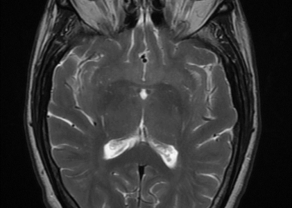
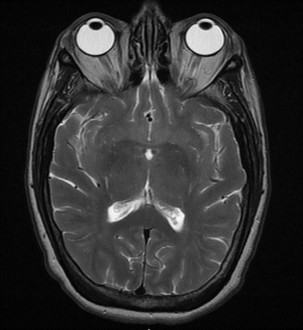
Orbits: There is no orbital mass, fluid collection or enhancing lesion. The optic nerves are symmetric and appear normal. The globes are intact. The rectus muscles are normal.
Brain: There is mild generalized atrophy. Areas of abnormal signal are seen within the periventricular and subcortical white matter, consistent with chronic small vessel ischemic disease. There is no cortical infarct, hemorrhage, mass or hydrocephalus.
Vasculature: Normal vascular flow voids are demonstrated. There is no evidence of arterial or venous occlusion.
Cavernous Sinuses: The cavernous sinus structures appear normal. There are small cysts along the left posterolateral margin of the pituitary gland, but these do not encroach on the contents of the cavernous sinuses.
Paranasal Sinuses: There are small mucous retention cysts in the maxillary sinuses.
IMPRESSION: Unremarkable appearance of the orbits. No mass or enhancing lesion.