


 MRI Case Study
Our case study of the month is an:
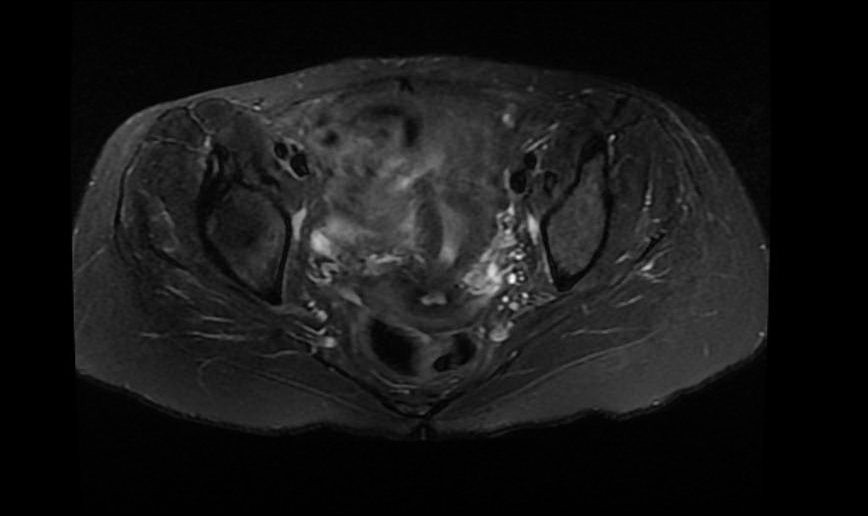
MRI of a Female Pelvis with and without IV Contrast
HISTORY: Lower abdominal and pelvic pain
TECHNIQUE: Multiplanar images of the pelvis were obtained at 1.5 Tesla prior to and following administration of 5 ml of Gadavist intravenous contrast.
FINDINGS:
Uterus: The uterus is normal in size and contour. The cervix also appears normal in size and contour. No cervical mass is seen. There are a few small incidental nabothian cysts within the cervix. There is a 6mm benign-appearing cyst along the right vaginal formix. There is a small amount of fluid in the cul-de-sac which is likely physiologic.
Endometrium: The endometrium is normal in thickness. The cervical canal appears normal.
Ovaries: The ovaries appear normal. No ovarian masses are seen. There are normal sized bilateral ovarian follicles.
Urinary Bladder: The urinary bladder appears normal, without stones or wall thickening.
Abdominal Wall: No abdominal wall or inguinal hernia is seen.
Lymphatics: No pelvic lymphadenopathy is present.
Bones: The bones appear unremarkable.
IMPRESSION: No evidence of cervical malignancy. No acute process. The ovaries are normal.