


MRI Foot Case Study
 Our case study of the month is an MRI scan of the left foot, status post motor vehicle accident. The 58 year old female patient presented with left foot pain in the lateral aspect near the malleolus and in the dorsal aspect of the distal metatarsals.The MRI Foot Case Study procedure included multi-planar images of the left foot obtained without contrast on our 1.5 Tesla MRI machine. There was no comparison scan.
MRI Exam Findings:
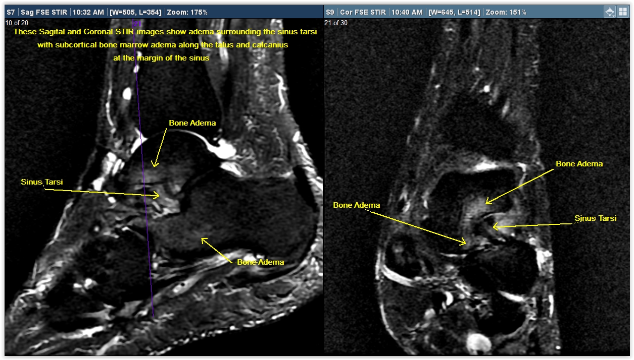
Bones: No fracture or dislocation is seen. There is mild bone marrow edema within the body of the talus, and within the subchondral bone of the calcaneus along the anterior subtalar facet. Findings may be associated with the sinus Tarsi syndrome. There are mild degenerative changes of the talonavicular joint. There are mild degenerative changes of the first metatarsophalangeal joint. There are small bone spurs along the posterior calcaneus at the Achilles’ tendon and plantar fascia insertions.
Tendons: The flexor and extensor tendons are intact. The Achilles tendon is intact. There is thickening and abnormal signal within the proximal fibers of the plantar fashion most likely related to chronic plantar fasciitis.
Ligaments: No evidence of ligament rupture is seen. The collateral ligaments are intact.
Soft Tissues: There is normal signal in the visualized soft tissues. No mass or abnormal fluid collection is seen. No significant joint effusion is present.
Impression: Bone marrow edema the talus and calcaneus likely related to sinus Tarsi syndrome. No fracture is seen. Chronic plantar fasciitis.
For more information on MRI imaging services at Greater Waterbury Imaging Center, visit our clinical section of the website.