


Our case study of the month is an:
MRI of the Lumbar Spine 3/28/2017
History: Lower Back Pain
 Comparison: MRI lumbar spine 7/30/2014
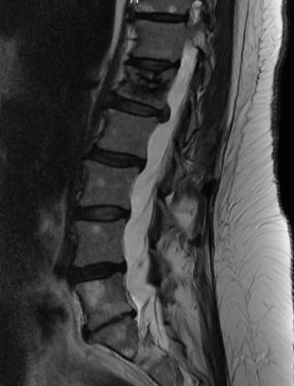
Technique: Axial and sagittal images of the lumbar spine were obtained at 1.5 Tesla.
Findings: The patient is status post L1 kyphoplasty. There is a chronic fracture along the superior endplate of L1. There is no acute fracture or subluxation. There is a benign hemangioma within the L5 vertebral body. There is no abnormal signal within the visible spinal cord. There are no intradural abnormalities. There is no paravertebral mass or fluid collection.
T12-L1: Normal. There is no disk herniation or evidence of neural compromise.
L1-L2: There is a small broad-based posterolateral disk protrusion with narrowing of the right neural foraminal ostium. The disk protrusion is similar in size compared to the previous study.
L2-L3: There is disk degeneration. There is a mild disk bulge without significant change. No focal disk herniation or evidence of neural compromise is seen.
L3-L4: There is mild diffuse disk bulge. There is no evidence of disk herniation or neural compromise.
L4-L5: There is mild disk degeneration. There is a minimal broad -based central disk protrusion. This is slightly increased from the prior study. There is no evidence of neural 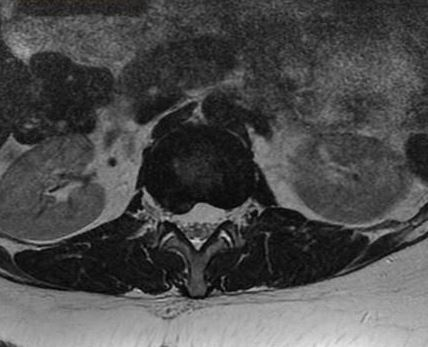 compromise.
L5-S1: There is disk degeneration with loss of disk height. There is a mild generalized disk bulge without evidence of disk herniation or neural compromise. The neural foramina are patent.
Impression: Small right posterolateral disk protrusion at L1-L2 without evidence of neural compromise at this level. No central disk protrusion at L4-L5, also without evidence of neural compromise.