


 Our case study of the month is an MRI of the head for a patient with a history of memory loss. The MRI Memory Loss Case Study procedure included multi-planar images of the head obtained without IV contrast on our 1.5 Tesla MRI machine. The comparison MRI scan was done on 1/18/2012.
History: Memory Loss
Technique: Multiplanar images of the head were obtained at 1.5 Tesla without IV contrast.
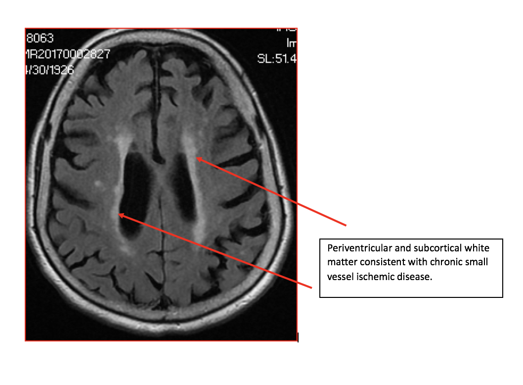
Brain: There is generalized atrophy. Areas of abnormal signal are seen within the periventricular and subcortical white matter consistent with chronic small vessel ischemic disease. These chronic white matter infarcts appear to have increased in number. There is no cortical infacrct, hemorrhage, mass or hydrocephalus.
Diffusion Images: No evidence of acute ischemic injury.
Pituitary: There is no sellar lesion.
IACS: The internal auditory canals are unremarkable.
Vasculature: Normal vascular flow voids are demonstrated.
Sinuses: There are small retention cysts or polyps in the maxillary sinuses.
IMPRESSION: No acute intracranial process. Atrophy with chronic small vessel ischemic disease. Small vessel ischemic disease has mildly progressed compared to the previous MRI from 2012.
For more information on MRI imaging services at Greater Waterbury Imaging Center, visit our clinical section of the website.